7RNI | pdb_00007rni

Discovery of an Anion-Dependent Farnesyltransferase Inhibitor from a Phenotypic Screen


Experimental Data Snapshot

  • Method: X-RAY DIFFRACTION
  • Resolution: 1.98 Å
  • R-Value Free: 
    0.185 (Depositor), 0.192 (DCC) 
  • R-Value Work: 
    0.162 (Depositor), 0.165 (DCC) 
  • R-Value Observed: 
    0.163 (Depositor) 

Starting Model: experimental
View more details

wwPDB Validation 3D Report Full Report

Validation slider image for 7RNI

Ligand Structure Quality Assessment 


This is version 1.1 of the entry. See complete history

Literature

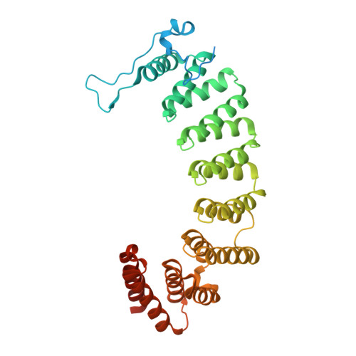
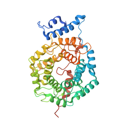
Discovery of an Anion-Dependent Farnesyltransferase Inhibitor from a Phenotypic Screen.

Bukhtiyarova, M.Cook, E.M.Hancock, P.J.Hruza, A.W.Shaw, A.W.Adam, G.C.Barnard, R.J.O.McKenna, P.M.Holloway, M.K.Bell, I.M.Carroll, S.Cornella-Taracido, I.Cox, C.D.Kutchukian, P.S.Powell, D.A.Strickland, C.Trotter, B.W.Tudor, M.Wolkenberg, S.Li, J.Tellers, D.M.

(2021) ACS Med Chem Lett 12: 99-106

  • DOI: https://doi.org/10.1021/acsmedchemlett.0c00551
  • Primary Citation Related Structures: 
    7RN5, 7RNI

  • PubMed Abstract: 

    By employing a phenotypic screen, a set of compounds, exemplified by 1 , were identified which potentiate the ability of histone deacetylase inhibitor vorinostat to reverse HIV latency. Proteome enrichment followed by quantitative mass spectrometric analysis employing a modified analogue of 1 as affinity bait identified farnesyl transferase (FTase) as the primary interacting protein in cell lysates. This ligand-FTase binding interaction was confirmed via X-ray crystallography and temperature dependent fluorescence studies, despite 1 lacking structural and binding similarity to known FTase inhibitors. Although multiple lines of evidence established the binding interaction, these ligands exhibited minimal inhibitory activity in a cell-free biochemical FTase inhibition assay. Subsequent modification of the biochemical assay by increasing anion concentration demonstrated FTase inhibitory activity in this novel class. We propose 1 binds together with the anion in the active site to inhibit farnesyl transferase. Implications for phenotypic screening deconvolution and HIV reactivation are discussed.


  • Organizational Affiliation
    • MRL, Merck & Co., Inc., West Point, Pennsylvania 19486, United States.

Macromolecule Content 

  • Total Structure Weight: 93.65 kDa 
  • Atom Count: 6,434 
  • Modeled Residue Count: 719 
  • Deposited Residue Count: 814 
  • Unique protein chains: 2

Macromolecules

Find similar proteins by:|  3D Structure
Entity ID: 1
MoleculeChains  Sequence LengthOrganismDetailsImage
Protein farnesyltransferase/geranylgeranyltransferase type-1 subunit alpha377Rattus norvegicusMutation(s): 0 
Gene Names: Fnta
EC: 2.5.1.58 (PDB Primary Data), 2.5.1.59 (PDB Primary Data)
UniProt
Find proteins for Q04631 (Rattus norvegicus)
Explore Q04631 
Go to UniProtKB:  Q04631
Entity Groups
Sequence Clusters30% Identity50% Identity70% Identity90% Identity95% Identity100% Identity
UniProt GroupQ04631
Sequence Annotations
Expand
Reference Sequence
Find similar proteins by:|  3D Structure
Entity ID: 2
MoleculeChains  Sequence LengthOrganismDetailsImage
Protein farnesyltransferase subunit beta437Rattus norvegicusMutation(s): 0 
Gene Names: Fntb
EC: 2.5.1.58
UniProt
Find proteins for Q02293 (Rattus norvegicus)
Explore Q02293 
Go to UniProtKB:  Q02293
Entity Groups
Sequence Clusters30% Identity50% Identity70% Identity90% Identity95% Identity100% Identity
UniProt GroupQ02293
Sequence Annotations
Expand
Reference Sequence

Small Molecules

Ligands 5 Unique
IDChains Name / Formula / InChI Key2D Diagram3D Interactions
60I
(Subject of Investigation/LOI)

Query on 60I


Download:Ideal Coordinates CCD File
E [auth B][4-[[3-(2,2-dimethylpropyl)-1~{H}-benzimidazol-2-yl]methyl]piperazin-1-yl]-(1,3,5-trimethylpyrazol-4-yl)methanone
C24 H34 N6 O
YHWDDYZFKUHHMW-UHFFFAOYSA-N
DMA

Query on DMA


Download:Ideal Coordinates CCD File
H [auth B]DIMETHYLALLYL DIPHOSPHATE
C5 H12 O7 P2
CBIDRCWHNCKSTO-UHFFFAOYSA-N
ZN

Query on ZN


Download:Ideal Coordinates CCD File
F [auth B]ZINC ION
Zn
PTFCDOFLOPIGGS-UHFFFAOYSA-N
ACE

Query on ACE


Download:Ideal Coordinates CCD File
C [auth A],
D [auth B]
ACETYL GROUP
C2 H4 O
IKHGUXGNUITLKF-UHFFFAOYSA-N
MG

Query on MG


Download:Ideal Coordinates CCD File
G [auth B]MAGNESIUM ION
Mg
JLVVSXFLKOJNIY-UHFFFAOYSA-N
Binding Affinity Annotations 
IDSourceBinding Affinity
60I BindingDB:  7RNI Kd: min: 340, max: 475 (nM) from 2 assay(s)

Experimental Data & Validation

Experimental Data

  • Method: X-RAY DIFFRACTION
  • Resolution: 1.98 Å
  • R-Value Free:  0.185 (Depositor), 0.192 (DCC) 
  • R-Value Work:  0.162 (Depositor), 0.165 (DCC) 
  • R-Value Observed: 0.163 (Depositor) 
Space Group: P 61
Unit Cell:
Length ( Å )Angle ( ˚ )
a = 169.058α = 90
b = 169.058β = 90
c = 68.846γ = 120
Software Package:
Software NamePurpose
BUSTERrefinement
autoPROCdata reduction
XDSdata reduction
autoPROCdata scaling
Aimlessdata scaling
MOLREPphasing

Structure Validation

View Full Validation Report


Ligand Structure Quality Assessment 


Entry History 

Deposition Data

Revision History  (Full details and data files)

  • Version 1.0: 2021-09-08
    Type: Initial release
  • Version 1.1: 2023-10-18
    Changes: Data collection, Refinement description