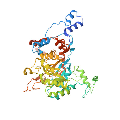
7RFI

HUMAN RETINAL VARIANT IMPDH1(595) TREATED WITH GTP, ATP, IMP, NAD+, INTERFACE-CENTERED

  • Classification: OXIDOREDUCTASE
  • Organism(s): Homo sapiens
  • Expression System: Escherichia coli BL21(DE3)
  • Mutation(s): No 

  • Deposited: 2021-07-14 Released: 2022-01-12 
  • Deposition Author(s): Burrell, A.L., Kollman, J.M.
  • Funding Organization(s): National Institutes of Health/National Institute of General Medical Sciences (NIH/NIGMS), National Institutes of Health/National Eye Institute (NIH/NEI)

Experimental Data Snapshot

  • Method: ELECTRON MICROSCOPY
  • Resolution: 2.60 Å
  • Aggregation State: FILAMENT 
  • Reconstruction Method: SINGLE PARTICLE 

wwPDB Validation   3D Report Full Report


This is version 1.3 of the entry. See complete history


Literature

IMPDH1 retinal variants control filament architecture to tune allosteric regulation.

Burrell, A.L.Nie, C.Said, M.Simonet, J.C.Fernandez-Justel, D.Johnson, M.C.Quispe, J.Buey, R.M.Peterson, J.R.Kollman, J.M.

(2022) Nat Struct Mol Biol 29: 47-58

  • DOI: https://doi.org/10.1038/s41594-021-00706-2
  • Primary Citation of Related Structures:  
    7RER, 7RES, 7RFE, 7RFF, 7RFG, 7RFH, 7RFI, 7RGD, 7RGI, 7RGL, 7RGM, 7RGQ

  • PubMed Abstract: 

    Inosine-5'-monophosphate dehydrogenase (IMPDH), a key regulatory enzyme in purine nucleotide biosynthesis, dynamically assembles filaments in response to changes in metabolic demand. Humans have two isoforms: IMPDH2 filaments reduce sensitivity to feedback inhibition, while IMPDH1 assembly remains uncharacterized. IMPDH1 plays a unique role in retinal metabolism, and point mutants cause blindness. Here, in a series of cryogenic-electron microscopy structures we show that human IMPDH1 assembles polymorphic filaments with different assembly interfaces in extended and compressed states. Retina-specific splice variants introduce structural elements that reduce sensitivity to GTP inhibition, including stabilization of the extended filament form. Finally, we show that IMPDH1 disease mutations fall into two classes: one disrupts GTP regulation and the other has no effect on GTP regulation or filament assembly. These findings provide a foundation for understanding the role of IMPDH1 in retinal function and disease and demonstrate the diverse mechanisms by which metabolic enzyme filaments are allosterically regulated.


  • Organizational Affiliation

    Department of Biochemistry, University of Washington, Seattle, WA, USA.


Macromolecules
Find similar proteins by:  (by identity cutoff)  |  3D Structure
Entity ID: 1
MoleculeChains Sequence LengthOrganismDetailsImage
Isoform 5 of Inosine-5'-monophosphate dehydrogenase 1
A, B, C, D, E
A, B, C, D, E, F, G, H
621Homo sapiensMutation(s): 0 
Gene Names: IMPDH1IMPD1
EC: 1.1.1.205
UniProt
Find proteins for Q5H9Q6 (Homo sapiens)
Explore Q5H9Q6 
Go to UniProtKB:  Q5H9Q6
Entity Groups  
Sequence Clusters30% Identity50% Identity70% Identity90% Identity95% Identity100% Identity
UniProt GroupQ5H9Q6
Sequence Annotations
Expand
  • Reference Sequence
Small Molecules
Ligands 3 Unique
IDChains Name / Formula / InChI Key2D Diagram3D Interactions
NAD (Subject of Investigation/LOI)
Query on NAD

Download Ideal Coordinates CCD File 
BA [auth E]
FA [auth F]
JA [auth G]
L [auth A]
NA [auth H]
BA [auth E],
FA [auth F],
JA [auth G],
L [auth A],
NA [auth H],
P [auth B],
T [auth C],
X [auth D]
NICOTINAMIDE-ADENINE-DINUCLEOTIDE
C21 H27 N7 O14 P2
BAWFJGJZGIEFAR-NNYOXOHSSA-N
GTP (Subject of Investigation/LOI)
Query on GTP

Download Ideal Coordinates CCD File 
CA [auth F]
DA [auth F]
GA [auth G]
HA [auth G]
I [auth A]
CA [auth F],
DA [auth F],
GA [auth G],
HA [auth G],
I [auth A],
J [auth A],
KA [auth H],
LA [auth H],
M [auth B],
N [auth B],
Q [auth C],
R [auth C],
U [auth D],
V [auth D],
Y [auth E],
Z [auth E]
GUANOSINE-5'-TRIPHOSPHATE
C10 H16 N5 O14 P3
XKMLYUALXHKNFT-UUOKFMHZSA-N
IMP (Subject of Investigation/LOI)
Query on IMP

Download Ideal Coordinates CCD File 
AA [auth E]
EA [auth F]
IA [auth G]
K [auth A]
MA [auth H]
AA [auth E],
EA [auth F],
IA [auth G],
K [auth A],
MA [auth H],
O [auth B],
S [auth C],
W [auth D]
INOSINIC ACID
C10 H13 N4 O8 P
GRSZFWQUAKGDAV-KQYNXXCUSA-N
Experimental Data & Validation

Experimental Data

  • Method: ELECTRON MICROSCOPY
  • Resolution: 2.60 Å
  • Aggregation State: FILAMENT 
  • Reconstruction Method: SINGLE PARTICLE 

Structure Validation

View Full Validation Report


Entry History & Funding Information

Deposition Data


Funding OrganizationLocationGrant Number
National Institutes of Health/National Institute of General Medical Sciences (NIH/NIGMS)United StatesR01GM118396
National Institutes of Health/National Eye Institute (NIH/NEI)United StatesEY030732
National Institutes of Health/National Institute of General Medical Sciences (NIH/NIGMS)United StatesGM008268

Revision History  (Full details and data files)

  • Version 1.0: 2022-01-12
    Type: Initial release
  • Version 1.1: 2022-01-26
    Changes: Database references
  • Version 1.2: 2022-02-02
    Changes: Database references
  • Version 1.3: 2024-06-05
    Changes: Data collection