


Crystal structure of the first bromodomain (BD1) of human BRD4 in complex with dual BRD4-JAK2 inhibitor MA9-060
Karim, M.R., Schonbrunn, E.To be published.
Experimental Data Snapshot
Starting Model: experimental
View more details
Entity ID: 1 | |||||
|---|---|---|---|---|---|
| Molecule | Chains | Sequence Length | Organism | Details | Image |
| Bromodomain-containing protein 4 | 127 | Homo sapiens | Mutation(s): 0 Gene Names: BRD4, HUNK1 | 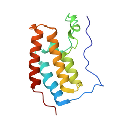 | |
UniProt & NIH Common Fund Data Resources | |||||
PHAROS: O60885 GTEx: ENSG00000141867 | |||||
Entity Groups | |||||
| Sequence Clusters | 30% Identity50% Identity70% Identity90% Identity95% Identity100% Identity | ||||
| UniProt Group | O60885 | ||||
Sequence AnnotationsExpand | |||||
Reference Sequence | |||||
| Ligands 2 Unique | |||||
|---|---|---|---|---|---|
| ID | Chains | Name / Formula / InChI Key | 2D Diagram | 3D Interactions | |
| 4OW (Subject of Investigation/LOI) Download:Ideal Coordinates CCD File | C [auth A] | N-[2-chloro-5-({2-[3-fluoro-4-(1-methylpiperidin-4-yl)anilino]-5-methylpyrimidin-4-yl}amino)phenyl]-2-methylpropane-2-sulfonamide C27 H34 Cl F N6 O2 S FPGNUYNSOYSDAN-UHFFFAOYSA-N |  | ||
| EDO Download:Ideal Coordinates CCD File | B [auth A] | 1,2-ETHANEDIOL C2 H6 O2 LYCAIKOWRPUZTN-UHFFFAOYSA-N |  | ||
| Length ( Å ) | Angle ( ˚ ) |
|---|---|
| a = 38.1 | α = 90 |
| b = 44.159 | β = 90 |
| c = 78.888 | γ = 90 |
| Software Name | Purpose |
|---|---|
| PHENIX | refinement |
| HKL-2000 | data scaling |
| PDB_EXTRACT | data extraction |
| HKL-2000 | data reduction |
| PHASER | phasing |
| Funding Organization | Location | Grant Number |
|---|---|---|
| Leukemia & Lymphoma Society | United States | -- |


