7RDF | pdb_00007rdf

Crystal structure of Pseudomonas aeruginosa D-Arginine Dehydrogenase Y249F co-crystallized in the presence of D-arginine


Experimental Data Snapshot

  • Method: X-RAY DIFFRACTION
  • Resolution: 1.29 Å
  • R-Value Free: 
    0.157 (Depositor), 0.157 (DCC) 
  • R-Value Work: 
    0.126 (Depositor), 0.128 (DCC) 
  • R-Value Observed: 
    0.128 (Depositor) 

Starting Model: experimental
View more details

wwPDB Validation 3D Report Full Report

Validation slider image for 7RDF

Ligand Structure Quality Assessment 


This is version 1.1 of the entry. See complete history

Literature

Discovery of a new flavin N5-adduct in a tyrosine to phenylalanine variant of d-Arginine dehydrogenase.

Iyer, A.Reis, R.A.G.Agniswamy, J.Weber, I.T.Gadda, G.

(2021) Arch Biochem Biophys 715: 109100-109100

  • DOI: https://doi.org/10.1016/j.abb.2021.109100
  • Primary Citation Related Structures: 
    7RDF

  • PubMed Abstract: 

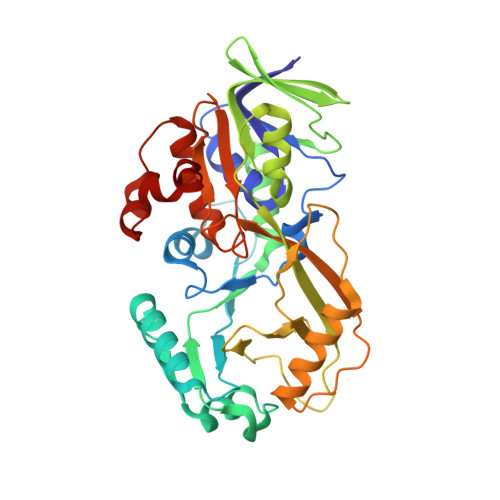
    d-Arginine dehydrogenase from Pseudomonas aeruginosa (PaDADH) catalyzes the flavin-dependent oxidation of d-arginine and other d-amino acids. Here, we report the crystal structure at 1.29 Å resolution for PaDADH-Y249F expressed and co-crystallized with d-arginine. The overall structure of PaDADH-Y249F resembled PaDADH-WT, but the electron density for the flavin cofactor was ambiguous, suggesting the presence of modified flavins. Electron density maps and mass spectrometric analysis confirmed the presence of both N5-(4-guanidino-oxobutyl)-FAD and 6-OH-FAD in a single crystal of PaDADH-Y249F and helped with the further refinement of the X-ray crystal structure. The versatility of the reduced flavin is apparent in the PaDADH-Y249F structure and is evidenced by the multiple functions it can perform in the same active site.


  • Organizational Affiliation
    • Department of Chemistry, Georgia State University, Atlanta, GA, 30302, USA.

Macromolecule Content 

  • Total Structure Weight: 42.69 kDa 
  • Atom Count: 3,816 
  • Modeled Residue Count: 375 
  • Deposited Residue Count: 375 
  • Unique protein chains: 1

Macromolecules

Find similar proteins by:|  3D Structure
Entity ID: 1
MoleculeChains  Sequence LengthOrganismDetailsImage
FAD-dependent catabolic D-arginine dehydrogenase DauA375Pseudomonas aeruginosa PAO1Mutation(s): 1 
Gene Names: dauAPA3863
EC: 1.4.99.6
UniProt
Find proteins for Q9HXE3 (Pseudomonas aeruginosa (strain ATCC 15692 / DSM 22644 / CIP 104116 / JCM 14847 / LMG 12228 / 1C / PRS 101 / PAO1))
Explore Q9HXE3 
Go to UniProtKB:  Q9HXE3
Entity Groups
Sequence Clusters30% Identity50% Identity70% Identity90% Identity95% Identity100% Identity
UniProt GroupQ9HXE3
Sequence Annotations
Expand
Reference Sequence

Small Molecules

Ligands 4 Unique
IDChains Name / Formula / InChI Key2D Diagram3D Interactions
4R2
(Subject of Investigation/LOI)

Query on 4R2


Download:Ideal Coordinates CCD File
B [auth A][[(2~{R},3~{S},4~{R},5~{R})-5-(6-aminopurin-9-yl)-3,4-bis(oxidanyl)oxolan-2-yl]methoxy-oxidanyl-phosphoryl] [(2~{R},3~{S},4~{S})-5-[5-[(~{E})-4-carbamimidamidobut-2-enoyl]-7,8-dimethyl-2,4-bis(oxidanylidene)-1~{H}-benzo[g]pteridin-10-yl]-2,3,4-tris(oxidanyl)pentyl] hydrogen phosphate
C32 H42 N12 O16 P2
UNPAMCKSCHHHHK-IGVXTPOKSA-N
6FA
(Subject of Investigation/LOI)

Query on 6FA


Download:Ideal Coordinates CCD File
C [auth A]6-HYDROXY-FLAVIN-ADENINE DINUCLEOTIDE
C27 H33 N9 O16 P2
BJSUUWFQAMLNKU-OKXKTURISA-N
PEG

Query on PEG


Download:Ideal Coordinates CCD File
G [auth A]DI(HYDROXYETHYL)ETHER
C4 H10 O3
MTHSVFCYNBDYFN-UHFFFAOYSA-N
GOL

Query on GOL


Download:Ideal Coordinates CCD File
D [auth A],
E [auth A],
F [auth A]
GLYCEROL
C3 H8 O3
PEDCQBHIVMGVHV-UHFFFAOYSA-N

Experimental Data & Validation

Experimental Data

  • Method: X-RAY DIFFRACTION
  • Resolution: 1.29 Å
  • R-Value Free:  0.157 (Depositor), 0.157 (DCC) 
  • R-Value Work:  0.126 (Depositor), 0.128 (DCC) 
  • R-Value Observed: 0.128 (Depositor) 
Space Group: P 21 21 21
Unit Cell:
Length ( Å )Angle ( ˚ )
a = 60.01α = 90
b = 76.33β = 90
c = 76.76γ = 90
Software Package:
Software NamePurpose
PHENIXrefinement
MOSFLMdata reduction
SCALAdata scaling
PHASERphasing

Structure Validation

View Full Validation Report


Ligand Structure Quality Assessment 


Entry History 

& Funding Information

Deposition Data


Funding OrganizationLocationGrant Number
National Science Foundation (NSF, United States)United StatesCHE1506518

Revision History  (Full details and data files)

  • Version 1.0: 2021-12-22
    Type: Initial release
  • Version 1.1: 2023-10-18
    Changes: Data collection, Refinement description