7RD8 | pdb_00007rd8

Structure of the S. cerevisiae P4B ATPase lipid flippase in the E1-ATP state


Experimental Data Snapshot

  • Method: ELECTRON MICROSCOPY
  • Resolution: 5.64 Å
  • Aggregation State: PARTICLE 
  • Reconstruction Method: SINGLE PARTICLE 

wwPDB Validation 3D Report Full Report

Validation slider image for 7RD8

This is version 1.3 of the entry. See complete history

Literature

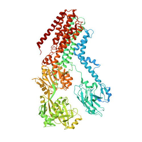
Structural basis of the P4B ATPase lipid flippase activity.

Bai, L.Jain, B.K.You, Q.Duan, H.D.Takar, M.Graham, T.R.Li, H.

(2021) Nat Commun 12: 5963-5963

  • DOI: https://doi.org/10.1038/s41467-021-26273-0
  • Primary Citation Related Structures: 
    7RD6, 7RD7, 7RD8

  • PubMed Abstract: 

    P4 ATPases are lipid flippases that are phylogenetically grouped into P4A, P4B and P4C clades. The P4A ATPases are heterodimers composed of a catalytic α-subunit and accessory β-subunit, and the structures of several heterodimeric flippases have been reported. The S. cerevisiae Neo1 and its orthologs represent the P4B ATPases, which function as monomeric flippases without a β-subunit. It has been unclear whether monomeric flippases retain the architecture and transport mechanism of the dimeric flippases. Here we report the structure of a P4B ATPase, Neo1, in its E1-ATP, E2P-transition, and E2P states. The structure reveals a conserved architecture as well as highly similar functional intermediate states relative to dimeric flippases. Consistently, structure-guided mutagenesis of residues in the proposed substrate translocation path disrupted Neo1's ability to establish membrane asymmetry. These observations indicate that evolutionarily distant P4 ATPases use a structurally conserved mechanism for substrate transport.


  • Organizational Affiliation
    • Department of Biochemistry and Biophysics, School of Basic Medical Sciences, Peking University, Beijing, China. lbai@bjmu.edu.cn.

Macromolecule Content 

  • Total Structure Weight: 130.89 kDa 
  • Atom Count: 7,417 
  • Modeled Residue Count: 929 
  • Deposited Residue Count: 1,151 
  • Unique protein chains: 1

Macromolecules

Find similar proteins by:|  3D Structure
Entity ID: 1
MoleculeChains  Sequence LengthOrganismDetailsImage
Probable phospholipid-transporting ATPase NEO11,151Saccharomyces cerevisiaeMutation(s): 0 
Gene Names: NEO1YIL048W
EC: 7.6.2.1
Membrane Entity: Yes 
UniProt
Find proteins for P40527 (Saccharomyces cerevisiae (strain ATCC 204508 / S288c))
Explore P40527 
Go to UniProtKB:  P40527
Entity Groups
Sequence Clusters30% Identity50% Identity70% Identity90% Identity95% Identity100% Identity
UniProt GroupP40527
Sequence Annotations
Expand
Reference Sequence

Small Molecules

Ligands 2 Unique
IDChains Name / Formula / InChI Key2D Diagram3D Interactions
ACP

Query on ACP


Download:Ideal Coordinates CCD File
B [auth A]PHOSPHOMETHYLPHOSPHONIC ACID ADENYLATE ESTER
C11 H18 N5 O12 P3
UFZTZBNSLXELAL-IOSLPCCCSA-N
MG

Query on MG


Download:Ideal Coordinates CCD File
C [auth A]MAGNESIUM ION
Mg
JLVVSXFLKOJNIY-UHFFFAOYSA-N

Experimental Data & Validation

Experimental Data

  • Method: ELECTRON MICROSCOPY
  • Resolution: 5.64 Å
  • Aggregation State: PARTICLE 
  • Reconstruction Method: SINGLE PARTICLE 
EM Software:
TaskSoftware PackageVersion
MODEL REFINEMENTPHENIX

Structure Validation

View Full Validation Report


Entry History 

& Funding Information

Deposition Data


Funding OrganizationLocationGrant Number
National Institutes of Health/National Institute of General Medical Sciences (NIH/NIGMS)United StatesCA231466 to H.L. and GM107978 to T.R.G.

Revision History  (Full details and data files)

  • Version 1.0: 2021-09-29
    Type: Initial release
  • Version 1.1: 2021-10-27
    Changes: Database references
  • Version 1.2: 2024-06-05
    Changes: Data collection
  • Version 1.3: 2025-05-28
    Changes: Data collection, Structure summary