7R6B

Crystal structure of mutant R43D/L124D/R125A/C273S of L-Asparaginase I from Yersinia pestis


Experimental Data Snapshot

  • Method: X-RAY DIFFRACTION
  • Resolution: 2.03 Å
  • R-Value Free: 0.218 
  • R-Value Work: 0.170 
  • R-Value Observed: 0.172 

wwPDB Validation   3D Report Full Report


This is version 1.3 of the entry. See complete history


Literature

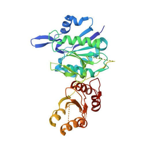
The dimeric form of bacterial l-asparaginase YpAI is fully active.

Strzelczyk, P.Zhang, D.Alexandratos, J.Piszczek, G.Wlodawer, A.Lubkowski, J.

(2023) FEBS J 290: 780-795

  • DOI: https://doi.org/10.1111/febs.16635
  • Primary Citation of Related Structures:  
    7R69, 7R6A, 7R6B

  • PubMed Abstract: 

    l-asparaginases from mesophilic bacteria (ASNases), including two enzymes very successfully used in the treatment of leukaemia, have been consistently described as homotetramers. On the contrary, structural studies show that homodimers of these enzymes should be sufficient to carry out the catalytic reaction. In this report, we investigated whether the type I Yersinia pestis asparaginase (YpAI) is active in a dimeric form or whether the tetrameric quaternary structure is critical for its activity. Using multiple biophysical techniques that investigate enzymatic properties and quaternary structure at either high or low protein concentration, we found that dimeric YpAI is fully active, suggesting that the tetrameric form of this subfamily of enzymes does not bear significant enzymatic relevance. In this process, we extensively characterized YpAI, showing that it is a cooperative enzyme, although the mechanism of allostery is still not definitely established. We showed that, like most type I ASNases, the substrate affinity of YpAI is low and this enzyme is very similar in terms of both the structure and enzymatic properties to homologous type I ASNase from Escherichia coli (EcAI). We extended these studies to more medically relevant type II ASNases, used as anti-leukaemia drugs. We confirmed that type II ASNases are not allosteric, and that they might also be functional in a dimeric form. However, the determination of the accurate tetramer⇆dimer dissociation constants of these enzymes that most likely lie in the picomolar range is not possible with currently available biophysical techniques.


  • Organizational Affiliation

    Center for Structural Biology, Center for Cancer Research, National Cancer Institute, Frederick, MD, USA.


Macromolecules
Find similar proteins by:  (by identity cutoff)  |  3D Structure
Entity ID: 1
MoleculeChains Sequence LengthOrganismDetailsImage
L-asparaginase I
A, B
338Yersinia pestisMutation(s): 4 
Gene Names: ansAYPO2161
EC: 3.5.1.1
UniProt
Find proteins for A0A3N4B0Q2 (Yersinia pestis)
Explore A0A3N4B0Q2 
Go to UniProtKB:  A0A3N4B0Q2
Entity Groups  
Sequence Clusters30% Identity50% Identity70% Identity90% Identity95% Identity100% Identity
UniProt GroupA0A3N4B0Q2
Sequence Annotations
Expand
  • Reference Sequence
Small Molecules
Ligands 2 Unique
IDChains Name / Formula / InChI Key2D Diagram3D Interactions
EDO
Query on EDO

Download Ideal Coordinates CCD File 
C [auth A]
D [auth A]
E [auth A]
F [auth A]
G [auth B]
C [auth A],
D [auth A],
E [auth A],
F [auth A],
G [auth B],
H [auth B],
I [auth B],
J [auth B],
K [auth B],
L [auth B]
1,2-ETHANEDIOL
C2 H6 O2
LYCAIKOWRPUZTN-UHFFFAOYSA-N
CA
Query on CA

Download Ideal Coordinates CCD File 
M [auth B],
N [auth B]
CALCIUM ION
Ca
BHPQYMZQTOCNFJ-UHFFFAOYSA-N
Experimental Data & Validation

Experimental Data

  • Method: X-RAY DIFFRACTION
  • Resolution: 2.03 Å
  • R-Value Free: 0.218 
  • R-Value Work: 0.170 
  • R-Value Observed: 0.172 
  • Space Group: C 2 2 21
Unit Cell:
Length ( Å )Angle ( ˚ )
a = 100.263α = 90
b = 115.893β = 90
c = 127.548γ = 90
Software Package:
Software NamePurpose
HKL-3000data reduction
HKL-3000data scaling
PHASERphasing
REFMACrefinement
PDB_EXTRACTdata extraction

Structure Validation

View Full Validation Report


Entry History & Funding Information

Deposition Data


Funding OrganizationLocationGrant Number
National Institutes of Health/National Cancer Institute (NIH/NCI)United States--

Revision History  (Full details and data files)

  • Version 1.0: 2022-07-06
    Type: Initial release
  • Version 1.1: 2022-10-05
    Changes: Database references
  • Version 1.2: 2023-02-08
    Changes: Database references
  • Version 1.3: 2023-10-25
    Changes: Data collection, Refinement description