Download Mendeley
Functional role of a tethered domain as a naturally fused cognate partner is demonstrated in a three-domain copper nitrite reductase.
Petchyam, N., Mbouku, A., Eady, R.R., Hasnain, S.S., Antonyuk, S.V.(2026) IUCrJ 13: 498-513
- PubMed: 42290377 Search on PubMedSearch on PubMed Central
- DOI: https://doi.org/10.1107/S2052252526004549
- Primary Citation Related Structures:
7QQ2, 7R2U, 8QGF, 9FOM, 9FUH, 9FUI, 9FUJ, 9FUK - PubMed Abstract:
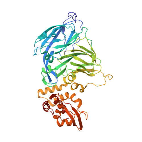
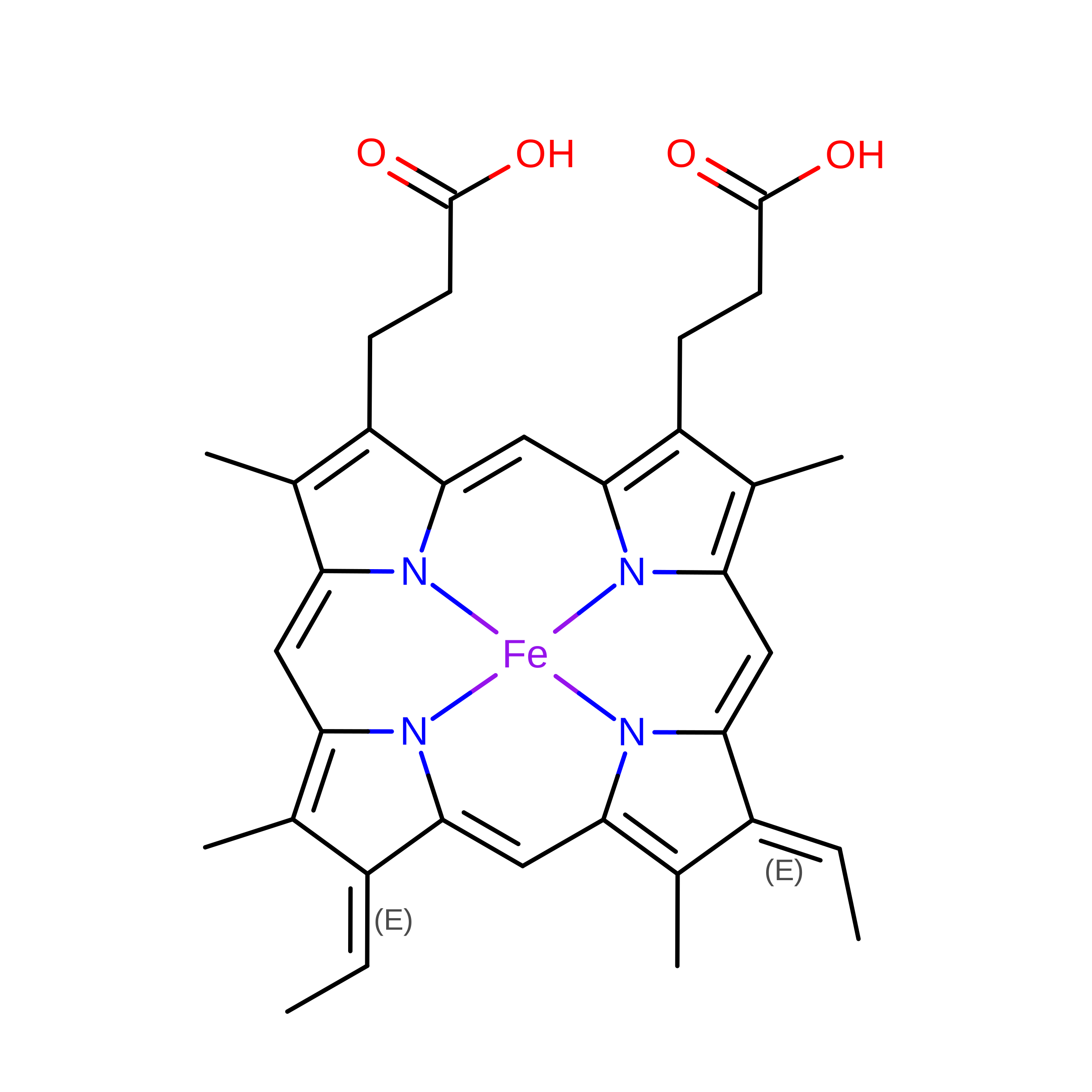
Copper-containing nitrite reductases (CuNiRs) catalyse the reduction of nitrite to nitric oxide and are a key enzyme in the anaerobic ammonium oxidation and denitrification steps of the nitrogen cycle. The recent recognition of the widespread distribution of three-domain CuNiRs where cognate redox partners are fused to the core NiR enzyme offered the possibility of studying coordinated events (e.g. proton-coupled electron transfer) in a conformationally stable donor-acceptor complex. The C-terminal cytochrome c tethered domain of the CuNiR from Ralstonia pickettii (RpNiR) has been well studied. Reverse engineering of RpNiR undertaken to remove the cognate partner domain showed that the presence of the additional domain resulted in significant differences in the apparent K m for nitrite and the reduction potentials of the Cu centres when compared with the core enzyme. The oxidation state of the haem centre and the position of the tethering linker have also been shown to control access of substrate to the active site. A key feature of this control is a conserved tyrosine residue (Tyr323 in RpNiR) located in the tethering linker between the fused domain and the core enzyme. To gain insight into this control, we have undertaken targeted mutations of RpNiR to probe the so-called primary proton channel and perturb putative electron transfer routes from the haem to the `gatekeeper' Tyr323 and to the T1Cu centre. The resolution of our crystallographic data to better than 1.2 Å enabled us to apply unrestrained SHELXL refinement of the structures. Our data provide a significant advance in our understanding of catalysis and modulation of electron transfer in these tethered systems, with wider implications for these fundamental processes in other protein complexes.
- Molecular Biophysics Group, Life Sciences Building, Institute of Systems, Molecular and Integrative Biology, Faculty of Health and Life Sciences, University of Liverpool, Liverpool L69 7ZB, United Kingdom.
Organizational Affiliation: