7R2M | pdb_00007r2m

SYNJ2BP complex with a synthetic Vangl2 peptide (9mer).


Experimental Data Snapshot

  • Method: X-RAY DIFFRACTION
  • Resolution: 2.40 Å
  • R-Value Free: 
    0.239 (Depositor), 0.240 (DCC) 
  • R-Value Work: 
    0.215 (Depositor), 0.215 (DCC) 
  • R-Value Observed: 
    0.216 (Depositor) 

Starting Model: experimental
View more details

wwPDB Validation   3D Report Full Report


Ligand Structure Quality Assessment 


This is version 1.2 of the entry. See complete history


Literature

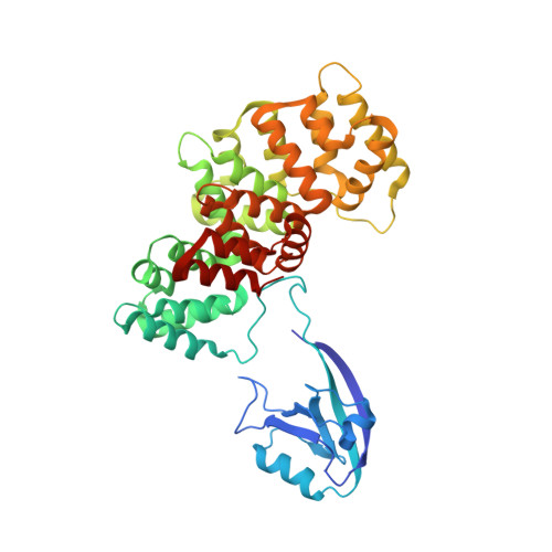
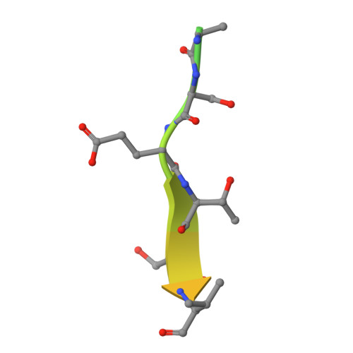
PDZome-wide and structural characterization of the PDZ-binding motif of VANGL2.

Montserrat-Gomez, M.Gogl, G.Carrasco, K.Betzi, S.Durbesson, F.Cousido-Siah, A.Kostmann, C.Essig, D.J.Stromgaard, K.Ostergaard, S.Morelli, X.Trave, G.Vincentelli, R.Bailly, E.Borg, J.P.

(2024) Biochim Biophys Acta Proteins Proteom 1872: 140989-140989

  • DOI: https://doi.org/10.1016/j.bbapap.2023.140989
  • Primary Citation of Related Structures:  
    7R2M, 7R2T

  • PubMed Abstract: 

    VANGL2 is a core component of the non-canonical Wnt/Planar Cell Polarity signaling pathway that uses its highly conserved carboxy-terminal type 1 PDZ-binding motif (PBM) to bind a variety of PDZ proteins. In this study, we characterize and quantitatively assess the largest VANGL2 PDZome-binding profile documented so far, using orthogonal methods. The results of our holdup approach support VANGL2 interactions with a large panel of both long-recognized and unprecedented PDZ domains. Truncation and point mutation analyses of the VANGL2 PBM establish that, beyond the strict requirement of the P-0 / V521 and P-2 / T519 amino acids, upstream residues, including E518, Q516 and R514 at, respectively, P-3, P-5 and P-7 further contribute to the robustness of VANGL2 interactions with two distinct PDZ domains, SNX27 and SCRIBBLE-PDZ3. In agreement with these data, incremental amino-terminal deletions of the VANGL2 PBM causes its overall affinity to progressively decline. Moreover, the holdup data establish that the PDZome binding repertoire of VANGL2 starts to diverge significantly with the truncation of E518. A structural analysis of the SYNJ2BP-PDZ/VANGL2 interaction with truncated PBMs identifies a major conformational change in the binding direction of the PBM peptide after the P-2 position. Finally, we report that the PDZome binding profile of VANGL2 is dramatically rearranged upon phosphorylation of S517, T519 and S520. Our crystallographic approach illustrates how SYNJ2BP accommodates a S520-phosphorylated PBM peptide through the ideal positioning of two basic residues, K48 and R86. Altogether our data provides a comprehensive view of the VANGL2 PDZ network and how this network specifically responds to the post-translation modification of distinct PBM residues. These findings should prove useful in guiding future functional and molecular studies of the key PCP component VANGL2.


  • Organizational Affiliation
    • Aix Marseille Université, CNRS, INSERM, Institut Paoli-Calmettes, CRCM, Equipe labellisée Ligue 'Cell polarity, cell signaling and cancer', Marseille, France.

Macromolecules
Find similar proteins by:  (by identity cutoff)  |  3D Structure
Entity ID: 1
MoleculeChains Sequence LengthOrganismDetailsImage
Synaptojanin-2-binding protein,AnnexinA [auth D],
B [auth A]
422Homo sapiensMutation(s): 0 
Gene Names: SYNJ2BPOMP25ANXA2
UniProt & NIH Common Fund Data Resources
Find proteins for P57105 (Homo sapiens)
Explore P57105 
Go to UniProtKB:  P57105
PHAROS:  P57105
GTEx:  ENSG00000213463 
Entity Groups  
Sequence Clusters30% Identity50% Identity70% Identity90% Identity95% Identity100% Identity
UniProt GroupP57105
Sequence Annotations
Expand
  • Reference Sequence

Find similar proteins by:  Sequence   |   3D Structure  

Entity ID: 2
MoleculeChains Sequence LengthOrganismDetailsImage
Vangl2 peptideC [auth B],
D [auth E]
24Homo sapiensMutation(s): 0 
UniProt & NIH Common Fund Data Resources
Find proteins for Q9ULK5 (Homo sapiens)
Explore Q9ULK5 
Go to UniProtKB:  Q9ULK5
PHAROS:  Q9ULK5
GTEx:  ENSG00000162738 
Entity Groups  
Sequence Clusters30% Identity50% Identity70% Identity90% Identity95% Identity100% Identity
UniProt GroupQ9ULK5
Sequence Annotations
Expand
  • Reference Sequence
Small Molecules
Ligands 3 Unique
IDChains Name / Formula / InChI Key2D Diagram3D Interactions
PE4 (Subject of Investigation/LOI)
Query on PE4

Download Ideal Coordinates CCD File 
K [auth D]2-{2-[2-(2-{2-[2-(2-ETHOXY-ETHOXY)-ETHOXY]-ETHOXY}-ETHOXY)-ETHOXY]-ETHOXY}-ETHANOL
C16 H34 O8
PJWQOENWHPEPKI-UHFFFAOYSA-N
GOL
Query on GOL

Download Ideal Coordinates CCD File 
I [auth D],
J [auth D],
P [auth A],
Q [auth A]
GLYCEROL
C3 H8 O3
PEDCQBHIVMGVHV-UHFFFAOYSA-N
CA
Query on CA

Download Ideal Coordinates CCD File 
E [auth D]
F [auth D]
G [auth D]
H [auth D]
L [auth D]
E [auth D],
F [auth D],
G [auth D],
H [auth D],
L [auth D],
M [auth A],
N [auth A],
O [auth A],
R [auth A]
CALCIUM ION
Ca
BHPQYMZQTOCNFJ-UHFFFAOYSA-N
Experimental Data & Validation

Experimental Data

  • Method: X-RAY DIFFRACTION
  • Resolution: 2.40 Å
  • R-Value Free:  0.239 (Depositor), 0.240 (DCC) 
  • R-Value Work:  0.215 (Depositor), 0.215 (DCC) 
  • R-Value Observed: 0.216 (Depositor) 
Space Group: P 1 21 1
Unit Cell:
Length ( Å )Angle ( ˚ )
a = 60.27α = 90
b = 60.71β = 92.88
c = 143.15γ = 90
Software Package:
Software NamePurpose
XSCALEdata scaling
PHENIXrefinement
PDB_EXTRACTdata extraction
XDSdata reduction
PHENIXphasing

Structure Validation

View Full Validation Report


Ligand Structure Quality Assessment 


Entry History & Funding Information

Deposition Data


Funding OrganizationLocationGrant Number
Not funded--

Revision History  (Full details and data files)

  • Version 1.0: 2023-05-17
    Type: Initial release
  • Version 1.1: 2024-02-07
    Changes: Data collection, Refinement description
  • Version 1.2: 2024-09-04
    Changes: Database references