7R0J | pdb_00007r0j

Structure of the V2 receptor Cter-arrestin2-ScFv30 complex


Experimental Data Snapshot

  • Method: ELECTRON MICROSCOPY
  • Resolution: 4.23 Å
  • Aggregation State: PARTICLE 
  • Reconstruction Method: SINGLE PARTICLE 

Starting Model: experimental
View more details

wwPDB Validation 3D Report Full Report

Validation slider image for 7R0J

This is version 1.2 of the entry. See complete history

Literature

Structure of the vasopressin hormone-V2 receptor-beta-arrestin1 ternary complex.

Bous, J.Fouillen, A.Orcel, H.Trapani, S.Cong, X.Fontanel, S.Saint-Paul, J.Lai-Kee-Him, J.Urbach, S.Sibille, N.Sounier, R.Granier, S.Mouillac, B.Bron, P.

(2022) Sci Adv 8: eabo7761-eabo7761

  • DOI: https://doi.org/10.1126/sciadv.abo7761
  • Primary Citation Related Structures: 
    7R0C, 7R0J

  • PubMed Abstract: 

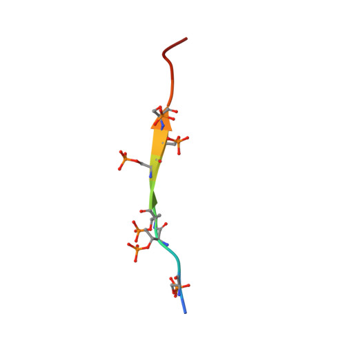
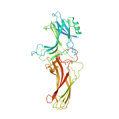
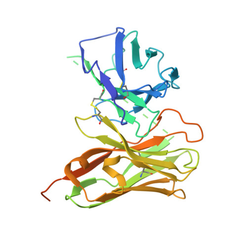
    Arrestins interact with G protein-coupled receptors (GPCRs) to stop G protein activation and to initiate key signaling pathways. Recent structural studies shed light on the molecular mechanisms involved in GPCR-arrestin coupling, but whether this process is conserved among GPCRs is poorly understood. Here, we report the cryo-electron microscopy active structure of the wild-type arginine-vasopressin V2 receptor (V2R) in complex with β-arrestin1. It reveals an atypical position of β-arrestin1 compared to previously described GPCR-arrestin assemblies, associated with an original V2R/β-arrestin1 interface involving all receptor intracellular loops. Phosphorylated sites of the V2R carboxyl terminus are clearly identified and interact extensively with the β-arrestin1 N-lobe, in agreement with structural data obtained with chimeric or synthetic systems. Overall, these findings highlight a notable structural variability among GPCR-arrestin signaling complexes.


  • Organizational Affiliation
    • CBS (Centre de Biologie Structurale), Université de Montpellier, CNRS, INSERM, Montpellier, France.

Macromolecule Content 

  • Total Structure Weight: 79.01 kDa 
  • Atom Count: 4,669 
  • Modeled Residue Count: 596 
  • Deposited Residue Count: 721 
  • Unique protein chains: 3

Macromolecules

Find similar proteins by:  Sequence   |   3D Structure  
Entity ID: 1
MoleculeChains  Sequence LengthOrganismDetailsImage
V2R Cter13Homo sapiensMutation(s): 0 
Membrane Entity: Yes 
UniProt & NIH Common Fund Data Resources
Find proteins for P30518 (Homo sapiens)
Explore P30518 
Go to UniProtKB:  P30518
PHAROS:  P30518
GTEx:  ENSG00000126895 
Entity Groups
Sequence Clusters30% Identity50% Identity70% Identity90% Identity95% Identity100% Identity
UniProt GroupP30518
Sequence Annotations
Expand
Reference Sequence
Find similar proteins by:|  3D Structure
Entity ID: 2
MoleculeChains  Sequence LengthOrganismDetailsImage
Arrestin2B [auth C]424Homo sapiensMutation(s): 0 
Membrane Entity: Yes 
UniProt & NIH Common Fund Data Resources
Find proteins for P49407 (Homo sapiens)
Explore P49407 
Go to UniProtKB:  P49407
PHAROS:  P49407
GTEx:  ENSG00000137486 
Entity Groups
Sequence Clusters30% Identity50% Identity70% Identity90% Identity95% Identity100% Identity
UniProt GroupP49407
Sequence Annotations
Expand
Reference Sequence
Find similar proteins by:|  3D Structure
Entity ID: 3
MoleculeChains  Sequence LengthOrganismDetailsImage
ScFv30C [auth D]284synthetic constructMutation(s): 0 

Small Molecules

Modified Residues  2 Unique
IDChains TypeFormula2D DiagramParent
SEP
Query on SEP
A
L-PEPTIDE LINKINGC3 H8 N O6 PSER
TPO
Query on TPO
A
L-PEPTIDE LINKINGC4 H10 N O6 PTHR

Experimental Data & Validation

Experimental Data

  • Method: ELECTRON MICROSCOPY
  • Resolution: 4.23 Å
  • Aggregation State: PARTICLE 
  • Reconstruction Method: SINGLE PARTICLE 
EM Software:
TaskSoftware PackageVersion
MODEL REFINEMENTPHENIX

Structure Validation

View Full Validation Report


Entry History 

& Funding Information

Deposition Data


Funding OrganizationLocationGrant Number
Agence Nationale de la Recherche (ANR)FranceANR-19-CE11-0014
Foundation for Medical Research (France)FranceDEQ20150331736

Revision History  (Full details and data files)

  • Version 1.0: 2022-09-14
    Type: Initial release
  • Version 1.1: 2024-10-16
    Changes: Data collection, Refinement description, Structure summary
  • Version 1.2: 2025-07-02
    Changes: Data collection