Unveiling molecular details behind improved activity at neutral to alkaline pH of an engineered DyP-type peroxidase.
Borges, P.T., Silva, D., Silva, T.F.D., Brissos, V., Canellas, M., Lucas, M.F., Masgrau, L., Melo, E.P., Machuqueiro, M., Frazao, C., Martins, L.O.(2022) Comput Struct Biotechnol J 20: 3899-3910
- PubMed: 35950185 Search on PubMedSearch on PubMed Central
- DOI: https://doi.org/10.1016/j.csbj.2022.07.032
- Primary Citation Related Structures:
7QYQ, 7QYZ, 7QZA - PubMed Abstract:
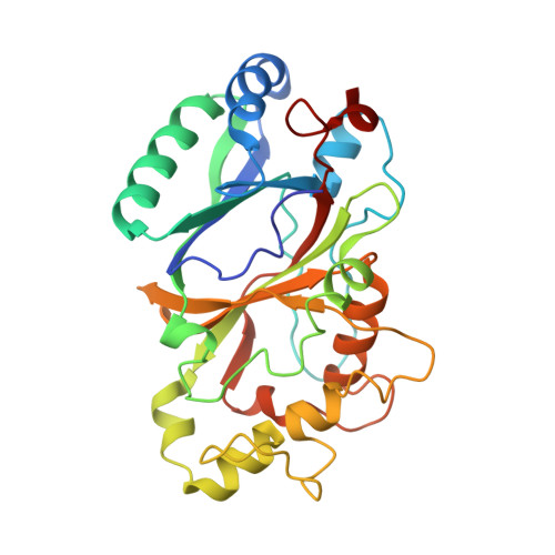
DyP-type peroxidases (DyPs) are microbial enzymes that catalyze the oxidation of a wide range of substrates, including synthetic dyes, lignin-derived compounds, and metals, such as Mn 2+ and Fe 2+ , and have enormous biotechnological potential in biorefineries. However, many questions on the molecular basis of enzyme function and stability remain unanswered. In this work, high-resolution structures of Pp DyP wild-type and two engineered variants (6E10 and 29E4) generated by directed evolution were obtained. The X-ray crystal structures revealed the typical ferredoxin-like folds, with three heme access pathways, two tunnels, and one cavity, limited by three long loops including catalytic residues. Variant 6E10 displays significantly increased loops' flexibility that favors function over stability: despite the considerably higher catalytic efficiency, this variant shows poorer protein stability compared to wild-type and 29E4 variants. Constant-pH MD simulations revealed a more positively charged microenvironment near the heme pocket of variant 6E10, particularly in the neutral to alkaline pH range. This microenvironment affects enzyme activity by modulating the p K a of essential residues in the heme vicinity and should account for variant 6E10 improved activity at pH 7-8 compared to the wild-type and 29E4 that show optimal enzymatic activity close to pH 4. Our findings shed light on the structure-function relationships of DyPs at the molecular level, including their pH-dependent conformational plasticity. These are essential for understanding and engineering the catalytic properties of DyPs for future biotechnological applications.
- Instituto de Tecnologia Química e Biológica António Xavier, Universidade NOVA de Lisboa, Av. da República, 2780-157 Oeiras, Portugal.
Organizational Affiliation: