


Structure of aminodeoxychorismate synthase component 1 (PabB) from Bacillus subtilis spizizenii.
Rooms, L.D., Race, P.R., Back, C.B., Burton, N.B., Willis, C.L., Stach, J.E.M., Duke, P.W., Hawkins, C.To be published.
Experimental Data Snapshot
Starting Model: experimental
View more details
Entity ID: 1 | |||||
|---|---|---|---|---|---|
| Molecule | Chains | Sequence Length | Organism | Details | Image |
| Anthranilate synthase component I family protein | 469 | Bacillus subtilis | Mutation(s): 1 Gene Names: FAL52_19525 | 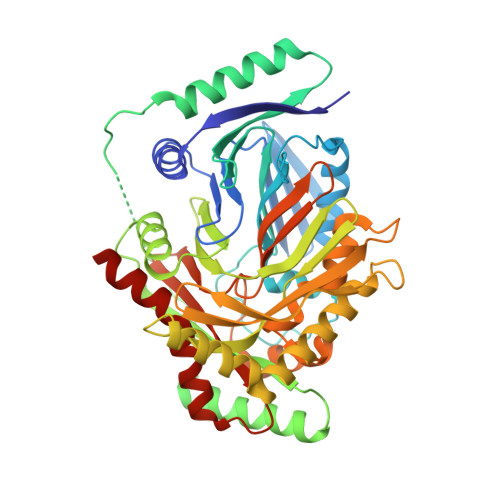 | |
| Ligands 2 Unique | |||||
|---|---|---|---|---|---|
| ID | Chains | Name / Formula / InChI Key | 2D Diagram | 3D Interactions | |
| TRP (Subject of Investigation/LOI) Download:Ideal Coordinates CCD File | F [auth A], G [auth B], H [auth C] | TRYPTOPHAN C11 H12 N2 O2 QIVBCDIJIAJPQS-VIFPVBQESA-N |  | ||
| GOL Download:Ideal Coordinates CCD File | D [auth A], E [auth A] | GLYCEROL C3 H8 O3 PEDCQBHIVMGVHV-UHFFFAOYSA-N |  | ||
| Length ( Å ) | Angle ( ˚ ) |
|---|---|
| a = 58.203 | α = 90 |
| b = 91.466 | β = 90.95 |
| c = 147.055 | γ = 90 |
| Software Name | Purpose |
|---|---|
| REFMAC | refinement |
| Aimless | data scaling |
| PDB_EXTRACT | data extraction |
| AutoProcess | data reduction |
| PHASER | phasing |
| Funding Organization | Location | Grant Number |
|---|---|---|
| Defence Science and Technology Laboratory (DSTL) | United Kingdom | -- |


