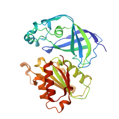
7QU3

X-ray structure of FAD domain of NqrF of Pseudomonas aeruginosa


Experimental Data Snapshot

  • Method: X-RAY DIFFRACTION
  • Resolution: 1.60 Å
  • R-Value Free: 0.196 
  • R-Value Work: 0.171 
  • R-Value Observed: 0.172 

wwPDB Validation   3D Report Full Report


Ligand Structure Quality Assessment 


This is version 1.2 of the entry. See complete history


Literature

Fast fragment- and compound-screening pipeline at the Swiss Light Source.

Kaminski, J.W.Vera, L.Stegmann, D.P.Vering, J.Eris, D.Smith, K.M.L.Huang, C.Y.Meier, N.Steuber, J.Wang, M.Fritz, G.Wojdyla, J.A.Sharpe, M.E.

(2022) Acta Crystallogr D Struct Biol 78: 328-336

  • DOI: https://doi.org/10.1107/S2059798322000705
  • Primary Citation of Related Structures:  
    7QTY, 7QU0, 7QU3, 7QU5

  • PubMed Abstract: 

    Over the last two decades, fragment-based drug discovery (FBDD) has emerged as an effective and efficient method to identify new chemical scaffolds for the development of lead compounds. X-ray crystallography can be used in FBDD as a tool to validate and develop fragments identified as binders by other methods. However, it is also often used with great success as a primary screening technique. In recent years, technological advances at macromolecular crystallography beamlines in terms of instrumentation, beam intensity and robotics have enabled the development of dedicated platforms at synchrotron sources for FBDD using X-ray crystallography. Here, the development of the Fast Fragment and Compound Screening (FFCS) platform, an integrated next-generation pipeline for crystal soaking, handling and data collection which allows crystallography-based screening of protein crystals against hundreds of fragments and compounds, at the Swiss Light Source is reported.


  • Organizational Affiliation

    Swiss Light Source, Paul Scherrer Institute, 5232 Villigen PSI, Switzerland.


Macromolecules
Find similar proteins by:  (by identity cutoff)  |  3D Structure
Entity ID: 1
MoleculeChains Sequence LengthOrganismDetailsImage
Na(+)-translocating NADH-quinone reductase subunit F280Pseudomonas aeruginosaMutation(s): 0 
Gene Names: nqrFPA14_25350
EC: 7.2.1.1
UniProt
Find proteins for Q02PF8 (Pseudomonas aeruginosa (strain UCBPP-PA14))
Explore Q02PF8 
Go to UniProtKB:  Q02PF8
Entity Groups  
Sequence Clusters30% Identity50% Identity70% Identity90% Identity95% Identity100% Identity
UniProt GroupQ02PF8
Sequence Annotations
Expand
  • Reference Sequence
Small Molecules
Ligands 4 Unique
IDChains Name / Formula / InChI Key2D Diagram3D Interactions
FAD
Query on FAD

Download Ideal Coordinates CCD File 
B [auth A]FLAVIN-ADENINE DINUCLEOTIDE
C27 H33 N9 O15 P2
VWWQXMAJTJZDQX-UYBVJOGSSA-N
RYM
Query on RYM

Download Ideal Coordinates CCD File 
C [auth A]4-(benzimidazol-1-ylmethyl)benzenecarbonitrile
C15 H11 N3
KZMYEQMVROHICO-UHFFFAOYSA-N
DMS
Query on DMS

Download Ideal Coordinates CCD File 
E [auth A],
F [auth A],
G [auth A],
H [auth A]
DIMETHYL SULFOXIDE
C2 H6 O S
IAZDPXIOMUYVGZ-UHFFFAOYSA-N
MG
Query on MG

Download Ideal Coordinates CCD File 
D [auth A]MAGNESIUM ION
Mg
JLVVSXFLKOJNIY-UHFFFAOYSA-N
Experimental Data & Validation

Experimental Data

  • Method: X-RAY DIFFRACTION
  • Resolution: 1.60 Å
  • R-Value Free: 0.196 
  • R-Value Work: 0.171 
  • R-Value Observed: 0.172 
  • Space Group: P 21 21 21
Unit Cell:
Length ( Å )Angle ( ˚ )
a = 49.543α = 90
b = 57.669β = 90
c = 101.44γ = 90
Software Package:
Software NamePurpose
PHENIXrefinement
PHENIXrefinement
XDSdata reduction
XSCALEdata scaling
PDB_EXTRACTdata extraction
PHASERphasing

Structure Validation

View Full Validation Report


Ligand Structure Quality Assessment 


Entry History & Funding Information

Deposition Data


Funding OrganizationLocationGrant Number
German Research Foundation (DFG)Germany--

Revision History  (Full details and data files)

  • Version 1.0: 2022-02-09
    Type: Initial release
  • Version 1.1: 2022-03-16
    Changes: Database references
  • Version 1.2: 2024-01-31
    Changes: Data collection, Refinement description