7QQF

Crystal structure of unliganded MYORG


Experimental Data Snapshot

  • Method: X-RAY DIFFRACTION
  • Resolution: 2.43 Å
  • R-Value Free: 0.233 
  • R-Value Work: 0.211 

wwPDB Validation   3D Report Full Report


Ligand Structure Quality Assessment 


This is version 1.2 of the entry. See complete history


Literature

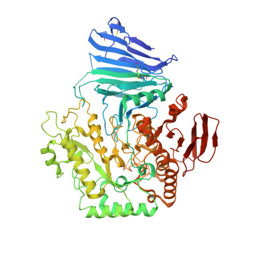
The primary familial brain calcification-associated protein MYORG is an alpha-galactosidase with restricted substrate specificity.

Meek, R.W.Brockerman, J.Fordwour, O.B.Zandberg, W.F.Davies, G.J.Vocadlo, D.J.

(2022) PLoS Biol 20: e3001764-e3001764

  • DOI: https://doi.org/10.1371/journal.pbio.3001764
  • Primary Citation of Related Structures:  
    7QQF, 7QQG, 7QQH

  • PubMed Abstract: 

    Primary familial brain calcification (PFBC) is characterised by abnormal deposits of calcium phosphate within various regions of the brain that are associated with severe cognitive impairments, psychiatric conditions, and movement disorders. Recent studies in diverse populations have shown a link between mutations in myogenesis-regulating glycosidase (MYORG) and the development of this disease. MYORG is a member of glycoside hydrolase (GH) family 31 (GH31) and, like the other mammalian GH31 enzyme α-glucosidase II, this enzyme is found in the lumen of the endoplasmic reticulum (ER). Though presumed to act as an α-glucosidase due to its localization and sequence relatedness to α-glucosidase II, MYORG has never been shown to exhibit catalytic activity. Here, we show that MYORG is an α-galactosidase and present the high-resolution crystal structure of MYORG in complex with substrate and inhibitor. Using these structures, we map detrimental mutations that are associated with MYORG-associated brain calcification and define how these mutations may drive disease progression through loss of enzymatic activity. Finally, we also detail the thermal stabilisation of MYORG afforded by a clinically approved small molecule ligand, opening the possibility of using pharmacological chaperones to enhance the activity of mutant forms of MYORG.


  • Organizational Affiliation

    Department of Chemistry. University of York, York, United Kingdom.


Macromolecules
Find similar proteins by:  (by identity cutoff)  |  3D Structure
Entity ID: 1
MoleculeChains Sequence LengthOrganismDetailsImage
Myogenesis-regulating glycosidase
A, B, C, D
636Homo sapiensMutation(s): 0 
Gene Names: MYORGKIAA1161
EC: 3.2.1
UniProt & NIH Common Fund Data Resources
Find proteins for Q6NSJ0 (Homo sapiens)
Explore Q6NSJ0 
Go to UniProtKB:  Q6NSJ0
PHAROS:  Q6NSJ0
GTEx:  ENSG00000164976 
Entity Groups  
Sequence Clusters30% Identity50% Identity70% Identity90% Identity95% Identity100% Identity
UniProt GroupQ6NSJ0
Sequence Annotations
Expand
  • Reference Sequence
Oligosaccharides

Help

Entity ID: 2
MoleculeChains Length2D Diagram Glycosylation3D Interactions
alpha-D-mannopyranose-(1-6)-beta-D-mannopyranose-(1-4)-2-acetamido-2-deoxy-beta-D-glucopyranose-(1-4)-2-acetamido-2-deoxy-beta-D-glucopyranose
E
4N-Glycosylation
Glycosylation Resources
GlyTouCan:  G22573RC
GlyCosmos:  G22573RC
GlyGen:  G22573RC
Entity ID: 3
MoleculeChains Length2D Diagram Glycosylation3D Interactions
2-acetamido-2-deoxy-beta-D-glucopyranose-(1-4)-2-acetamido-2-deoxy-beta-D-glucopyranose
F, G, H, K, L
F, G, H, K, L, M, N
2N-Glycosylation
Glycosylation Resources
GlyTouCan:  G42666HT
GlyCosmos:  G42666HT
GlyGen:  G42666HT
Entity ID: 4
MoleculeChains Length2D Diagram Glycosylation3D Interactions
beta-D-mannopyranose-(1-4)-2-acetamido-2-deoxy-beta-D-glucopyranose-(1-4)-2-acetamido-2-deoxy-beta-D-glucopyranose
I
3N-Glycosylation
Glycosylation Resources
GlyTouCan:  G15407YE
GlyCosmos:  G15407YE
GlyGen:  G15407YE
Entity ID: 5
MoleculeChains Length2D Diagram Glycosylation3D Interactions
alpha-L-fucopyranose-(1-3)-[2-acetamido-2-deoxy-beta-D-glucopyranose-(1-4)][alpha-L-fucopyranose-(1-6)]2-acetamido-2-deoxy-beta-D-glucopyranose
J
4N-Glycosylation
Glycosylation Resources
GlyTouCan:  G63564LA
GlyCosmos:  G63564LA
Small Molecules
Ligands 3 Unique
IDChains Name / Formula / InChI Key2D Diagram3D Interactions
NAG
Query on NAG

Download Ideal Coordinates CCD File 
AA [auth C]
DA [auth D]
EA [auth D]
O [auth A]
P [auth A]
AA [auth C],
DA [auth D],
EA [auth D],
O [auth A],
P [auth A],
Q [auth A],
T [auth B],
U [auth B],
V [auth B],
Y [auth C],
Z [auth C]
2-acetamido-2-deoxy-beta-D-glucopyranose
C8 H15 N O6
OVRNDRQMDRJTHS-FMDGEEDCSA-N
MLI
Query on MLI

Download Ideal Coordinates CCD File 
BA [auth C],
GA [auth D],
S [auth A],
W [auth B]
MALONATE ION
C3 H2 O4
OFOBLEOULBTSOW-UHFFFAOYSA-L
ACT
Query on ACT

Download Ideal Coordinates CCD File 
CA [auth C],
FA [auth D],
R [auth A],
X [auth B]
ACETATE ION
C2 H3 O2
QTBSBXVTEAMEQO-UHFFFAOYSA-M
Experimental Data & Validation

Experimental Data

  • Method: X-RAY DIFFRACTION
  • Resolution: 2.43 Å
  • R-Value Free: 0.233 
  • R-Value Work: 0.211 
  • Space Group: P 1
Unit Cell:
Length ( Å )Angle ( ˚ )
a = 74.109α = 88.062
b = 78.986β = 78.801
c = 180.113γ = 62.714
Software Package:
Software NamePurpose
REFMACrefinement
DIALSdata reduction
Aimlessdata scaling
PHASERphasing

Structure Validation

View Full Validation Report


Ligand Structure Quality Assessment 


Entry History & Funding Information

Deposition Data


Funding OrganizationLocationGrant Number
Royal SocietyUnited Kingdom180016

Revision History  (Full details and data files)

  • Version 1.0: 2022-08-31
    Type: Initial release
  • Version 1.1: 2023-01-18
    Changes: Database references
  • Version 1.2: 2024-01-31
    Changes: Data collection, Refinement description