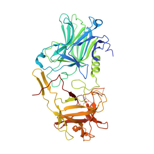
7QPT

Botulinum neurotoxin A4 cell binding domain in complex with GD1a oligosaccharide


Experimental Data Snapshot

  • Method: X-RAY DIFFRACTION
  • Resolution: 2.30 Å
  • R-Value Free: 0.247 
  • R-Value Work: 0.203 
  • R-Value Observed: 0.205 

wwPDB Validation   3D Report Full Report


This is version 1.1 of the entry. See complete history


Literature

Crystal Structures of Botulinum Neurotoxin Subtypes A4 and A5 Cell Binding Domains in Complex with Receptor Ganglioside.

Gregory, K.S.Mojanaga, O.O.Liu, S.M.Acharya, K.R.

(2022) Toxins (Basel) 14

  • DOI: https://doi.org/10.3390/toxins14020129
  • Primary Citation of Related Structures:  
    7QPT, 7QPU

  • PubMed Abstract: 

    Botulinum neurotoxins (BoNT) cause the potentially fatal neuroparalytic disease botulism that arises due to proteolysis of a SNARE protein. Each BoNT is comprised of three domains: a cell binding domain (H C ), a translocation domain (H N ), and a catalytic (Zn 2+ endopeptidase) domain (LC). The H C is responsible for neuronal specificity by targeting both a protein and ganglioside receptor at the neuromuscular junction. Although highly toxic, some BoNTs are commercially available as therapeutics for the treatment of a range of neuromuscular conditions. Here we present the crystal structures of two BoNT cell binding domains, H C /A4 and H C /A5, in a complex with the oligosaccharide of ganglioside, GD1a and GM1b, respectively. These structures, along with a detailed comparison with the previously reported apo-structures, reveal the conformational changes that occur upon ganglioside binding and the interactions involved.


  • Organizational Affiliation

    Department of Biology and Biochemistry, University of Bath, Claverton Down, Bath BA2 7AY, UK.


Macromolecules
Find similar proteins by:  (by identity cutoff)  |  3D Structure
Entity ID: 1
MoleculeChains Sequence LengthOrganismDetailsImage
Neurotoxin type A
A, B
433Clostridium botulinumMutation(s): 0 
UniProt
Find proteins for Q3LRX8 (Clostridium botulinum)
Explore Q3LRX8 
Go to UniProtKB:  Q3LRX8
Entity Groups  
Sequence Clusters30% Identity50% Identity70% Identity90% Identity95% Identity100% Identity
UniProt GroupQ3LRX8
Sequence Annotations
Expand
  • Reference Sequence
Oligosaccharides

Help

Entity ID: 2
MoleculeChains Length2D Diagram Glycosylation3D Interactions
N-acetyl-alpha-neuraminic acid-(2-3)-beta-D-galactopyranose-(1-3)-2-acetamido-2-deoxy-beta-D-galactopyranose-(1-4)-beta-D-galactopyranose-(1-4)-beta-D-glucopyranose
C, D
5N/A
Glycosylation Resources
GlyTouCan:  G03277YI
GlyCosmos:  G03277YI
GlyGen:  G03277YI
Experimental Data & Validation

Experimental Data

  • Method: X-RAY DIFFRACTION
  • Resolution: 2.30 Å
  • R-Value Free: 0.247 
  • R-Value Work: 0.203 
  • R-Value Observed: 0.205 
  • Space Group: P 61
Unit Cell:
Length ( Å )Angle ( ˚ )
a = 94.677α = 90
b = 94.677β = 90
c = 181.209γ = 120
Software Package:
Software NamePurpose
REFMACrefinement
PHENIXrefinement
DIALSdata reduction
Aimlessdata scaling
PHASERphasing

Structure Validation

View Full Validation Report


Entry History & Funding Information

Deposition Data


Funding OrganizationLocationGrant Number
Not funded--

Revision History  (Full details and data files)

  • Version 1.0: 2022-03-16
    Type: Initial release
  • Version 1.1: 2024-01-31
    Changes: Data collection, Refinement description