7QOA | pdb_00007qoa

Structure of CodB, a cytosine transporter in an outward-facing conformation


Experimental Data Snapshot

  • Method: X-RAY DIFFRACTION
  • Resolution: 2.40 Å
  • R-Value Free: 
    0.265 (Depositor), 0.270 (DCC) 
  • R-Value Work: 
    0.218 (Depositor) 
  • R-Value Observed: 
    0.220 (Depositor) 

Starting Model: experimental
View more details

wwPDB Validation 3D Report Full Report

Validation slider image for 7QOA

Ligand Structure Quality Assessment 


This is version 1.2 of the entry. See complete history

Literature

Structure of cytosine transport protein CodB provides insight into nucleobase-cation symporter 1 mechanism.

Hatton, C.E.Brotherton, D.H.Spencer, M.Cameron, A.D.

(2022) EMBO J 41: e110527-e110527

  • DOI: https://doi.org/10.15252/embj.2021110527
  • Primary Citation Related Structures: 
    7QOA

  • PubMed Abstract: 

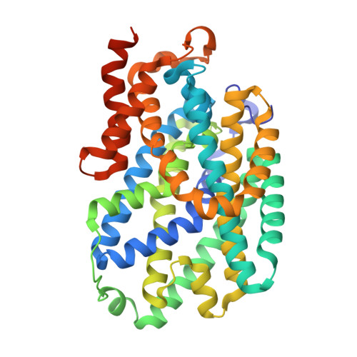
    CodB is a cytosine transporter from the Nucleobase-Cation-Symport-1 (NCS1) transporter family, a member of the widespread LeuT superfamily. Previous experiments with the nosocomial pathogen Pseudomonas aeruginosa have shown CodB as also important for the uptake of 5-fluorocytosine, which has been suggested as a novel drug to combat antimicrobial resistance by suppressing virulence. Here we solve the crystal structure of CodB from Proteus vulgaris, at 2.4 Å resolution in complex with cytosine. We show that CodB carries out the sodium-dependent uptake of cytosine and can bind 5-fluorocytosine. Comparison of the substrate-bound structures of CodB and the hydantoin transporter Mhp1, the only other NCS1 family member for which the structure is known, highlight the importance of the hydrogen bonds that the substrates make with the main chain at the breakpoint in the discontinuous helix, TM6. In contrast to other LeuT superfamily members, neither CodB nor Mhp1 makes specific interactions with residues on TM1. Comparison of the structures provides insight into the intricate mechanisms of how these proteins transport substrates across the plasma membrane.


  • Organizational Affiliation
    • School of Life Sciences, University of Warwick, Coventry, UK.

Macromolecule Content 

  • Total Structure Weight: 91.64 kDa 
  • Atom Count: 6,083 
  • Modeled Residue Count: 801 
  • Deposited Residue Count: 846 
  • Unique protein chains: 1

Macromolecules

Find similar proteins by:|  3D Structure
Entity ID: 1
MoleculeChains  Sequence LengthOrganismDetailsImage
Cytosine permease
A, B
423Proteus vulgarisMutation(s): 0 
Gene Names: EKQ45_12260

Small Molecules

Ligands 5 Unique
IDChains Name / Formula / InChI Key2D Diagram3D Interactions
PEF

Query on PEF


Download:Ideal Coordinates CCD File
D [auth A]DI-PALMITOYL-3-SN-PHOSPHATIDYLETHANOLAMINE
C37 H74 N O8 P
SLKDGVPOSSLUAI-PGUFJCEWSA-N
LMT

Query on LMT


Download:Ideal Coordinates CCD File
E [auth A]DODECYL-BETA-D-MALTOSIDE
C24 H46 O11
NLEBIOOXCVAHBD-QKMCSOCLSA-N
A6L

Query on A6L


Download:Ideal Coordinates CCD File
F [auth A],
G [auth A],
J [auth B],
K [auth B],
L [auth B]
2,3-dihydroxypropyl (9Z)-octadec-9-enoate
C21 H40 O4
RZRNAYUHWVFMIP-KTKRTIGZSA-N
CYT
(Subject of Investigation/LOI)

Query on CYT


Download:Ideal Coordinates CCD File
C [auth A],
I [auth B]
6-AMINOPYRIMIDIN-2(1H)-ONE
C4 H5 N3 O
OPTASPLRGRRNAP-UHFFFAOYSA-N
NA
(Subject of Investigation/LOI)

Query on NA


Download:Ideal Coordinates CCD File
H [auth A],
M [auth B]
SODIUM ION
Na
FKNQFGJONOIPTF-UHFFFAOYSA-N

Experimental Data & Validation

Experimental Data

  • Method: X-RAY DIFFRACTION
  • Resolution: 2.40 Å
  • R-Value Free:  0.265 (Depositor), 0.270 (DCC) 
  • R-Value Work:  0.218 (Depositor) 
  • R-Value Observed: 0.220 (Depositor) 
Space Group: C 2 2 21
Unit Cell:
Length ( Å )Angle ( ˚ )
a = 108.246α = 90
b = 209.027β = 90
c = 102.452γ = 90
Software Package:
Software NamePurpose
PHENIXrefinement
PHENIXrefinement
DIALSdata reduction
Aimlessdata scaling
MR-Rosettaphasing

Structure Validation

View Full Validation Report


Ligand Structure Quality Assessment 


Entry History 

& Funding Information

Deposition Data


Funding OrganizationLocationGrant Number
Biotechnology and Biological Sciences Research Council (BBSRC)United Kingdom--

Revision History  (Full details and data files)

  • Version 1.0: 2022-07-13
    Type: Initial release
  • Version 1.1: 2022-08-24
    Changes: Database references
  • Version 1.2: 2024-01-31
    Changes: Data collection, Refinement description