7QL5

Torpedo muscle-type nicotinic acetylcholine receptor - nicotine-bound conformation


Experimental Data Snapshot

  • Method: ELECTRON MICROSCOPY
  • Resolution: 2.50 Å
  • Aggregation State: PARTICLE 
  • Reconstruction Method: SINGLE PARTICLE 

wwPDB Validation   3D Report Full Report


This is version 1.2 of the entry. See complete history


Literature

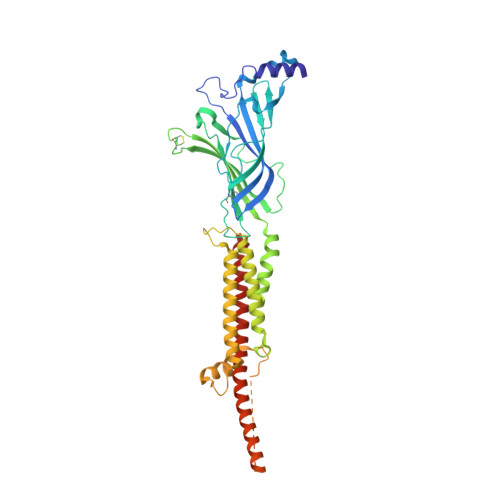
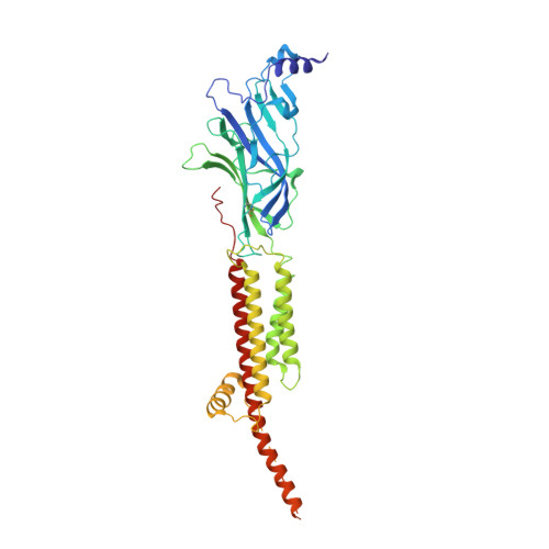
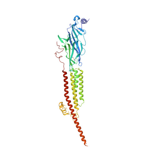
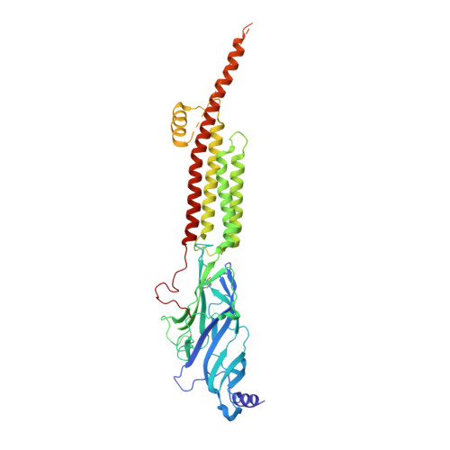
Conformational transitions and ligand-binding to a muscle-type nicotinic acetylcholine receptor.

Zarkadas, E.Pebay-Peyroula, E.Thompson, M.J.Schoehn, G.Uchanski, T.Steyaert, J.Chipot, C.Dehez, F.Baenziger, J.E.Nury, H.

(2022) Neuron 110: 1358

  • DOI: https://doi.org/10.1016/j.neuron.2022.01.013
  • Primary Citation of Related Structures:  
    7QKO, 7QL5, 7QL6

  • PubMed Abstract: 

    Fast synaptic communication requires receptors that respond to the presence of neurotransmitter by opening an ion channel across the post-synaptic membrane. The muscle-type nicotinic acetylcholine receptor from the electric fish, Torpedo, is the prototypic ligand-gated ion channel, yet the structural changes underlying channel activation remain undefined. Here we use cryo-EM to solve apo and agonist-bound structures of the Torpedo nicotinic receptor embedded in a lipid nanodisc. Using both a direct biochemical assay to define the conformational landscape and molecular dynamics simulations to assay flux through the pore, we correlate structures with functional states and elucidate the motions that lead to pore activation of a heteromeric nicotinic receptor. We highlight an underappreciated role for the complementary subunit in channel gating, establish the structural basis for the differential agonist affinities of α/δ versus α /γ sites, and explain why nicotine is less potent at muscle nicotinic receptors compared to neuronal ones.


  • Organizational Affiliation

    Université Grenoble Alpes, CNRS, CEA, IBS, F-38000 Grenoble, France.


Macromolecules
Find similar proteins by:  (by identity cutoff)  |  3D Structure
Entity ID: 1
MoleculeChains Sequence LengthOrganismDetailsImage
Acetylcholine receptor subunit alpha
A, D
437Tetronarce californicaMutation(s): 0 
Membrane Entity: Yes 
UniProt
Find proteins for P02710 (Tetronarce californica)
Explore P02710 
Go to UniProtKB:  P02710
Entity Groups  
Sequence Clusters30% Identity50% Identity70% Identity90% Identity95% Identity100% Identity
UniProt GroupP02710
Sequence Annotations
Expand
  • Reference Sequence
Find similar proteins by:  (by identity cutoff)  |  3D Structure
Entity ID: 2
MoleculeChains Sequence LengthOrganismDetailsImage
Acetylcholine receptor subunit beta469Tetronarce californicaMutation(s): 0 
Membrane Entity: Yes 
UniProt
Find proteins for P02712 (Tetronarce californica)
Explore P02712 
Go to UniProtKB:  P02712
Entity Groups  
Sequence Clusters30% Identity50% Identity70% Identity90% Identity95% Identity100% Identity
UniProt GroupP02712
Sequence Annotations
Expand
  • Reference Sequence
Find similar proteins by:  (by identity cutoff)  |  3D Structure
Entity ID: 3
MoleculeChains Sequence LengthOrganismDetailsImage
Acetylcholine receptor subunit delta501Tetronarce californicaMutation(s): 0 
Membrane Entity: Yes 
UniProt
Find proteins for P02718 (Tetronarce californica)
Explore P02718 
Go to UniProtKB:  P02718
Entity Groups  
Sequence Clusters30% Identity50% Identity70% Identity90% Identity95% Identity100% Identity
UniProt GroupP02718
Sequence Annotations
Expand
  • Reference Sequence
Find similar proteins by:  (by identity cutoff)  |  3D Structure
Entity ID: 4
MoleculeChains Sequence LengthOrganismDetailsImage
Acetylcholine receptor subunit gamma489Tetronarce californicaMutation(s): 0 
Membrane Entity: Yes 
UniProt
Find proteins for P02714 (Tetronarce californica)
Explore P02714 
Go to UniProtKB:  P02714
Entity Groups  
Sequence Clusters30% Identity50% Identity70% Identity90% Identity95% Identity100% Identity
UniProt GroupP02714
Sequence Annotations
Expand
  • Reference Sequence
Oligosaccharides

Help

Entity ID: 5
MoleculeChains Length2D Diagram Glycosylation3D Interactions
alpha-D-mannopyranose-(1-3)-[alpha-D-mannopyranose-(1-6)]alpha-D-mannopyranose-(1-6)-[alpha-D-mannopyranose-(1-3)]beta-D-mannopyranose-(1-4)-2-acetamido-2-deoxy-beta-D-glucopyranose-(1-4)-2-acetamido-2-deoxy-beta-D-glucopyranose
F
7N-Glycosylation
Glycosylation Resources
GlyTouCan:  G55220VL
GlyCosmos:  G55220VL
GlyGen:  G55220VL
Entity ID: 6
MoleculeChains Length2D Diagram Glycosylation3D Interactions
alpha-D-mannopyranose-(1-3)-[alpha-D-mannopyranose-(1-6)]beta-D-mannopyranose-(1-4)-2-acetamido-2-deoxy-beta-D-glucopyranose-(1-4)-2-acetamido-2-deoxy-beta-D-glucopyranose
G, H, I
5N-Glycosylation
Glycosylation Resources
GlyTouCan:  G22768VO
GlyCosmos:  G22768VO
GlyGen:  G22768VO
Entity ID: 7
MoleculeChains Length2D Diagram Glycosylation3D Interactions
2-acetamido-2-deoxy-beta-D-glucopyranose-(1-4)-2-acetamido-2-deoxy-beta-D-glucopyranose
J
2N-Glycosylation
Glycosylation Resources
GlyTouCan:  G42666HT
GlyCosmos:  G42666HT
GlyGen:  G42666HT
Entity ID: 8
MoleculeChains Length2D Diagram Glycosylation3D Interactions
alpha-D-mannopyranose-(1-6)-alpha-D-mannopyranose-(1-6)-[alpha-D-mannopyranose-(1-3)]beta-D-mannopyranose-(1-4)-2-acetamido-2-deoxy-beta-D-glucopyranose-(1-4)-2-acetamido-2-deoxy-beta-D-glucopyranose
K, L
6N-Glycosylation
Glycosylation Resources
GlyTouCan:  G34442SS
GlyCosmos:  G34442SS
GlyGen:  G34442SS
Small Molecules
Ligands 3 Unique
IDChains Name / Formula / InChI Key2D Diagram3D Interactions
POV (Subject of Investigation/LOI)
Query on POV

Download Ideal Coordinates CCD File 
N [auth A]
O [auth A]
P [auth A]
Q [auth B]
R [auth C]
N [auth A],
O [auth A],
P [auth A],
Q [auth B],
R [auth C],
S [auth C],
U [auth D],
V [auth D],
X [auth E],
Y [auth E],
Z [auth E]
(2S)-3-(hexadecanoyloxy)-2-[(9Z)-octadec-9-enoyloxy]propyl 2-(trimethylammonio)ethyl phosphate
C42 H82 N O8 P
WTJKGGKOPKCXLL-PFDVCBLKSA-N
NAG
Query on NAG

Download Ideal Coordinates CCD File 
W [auth E]2-acetamido-2-deoxy-beta-D-glucopyranose
C8 H15 N O6
OVRNDRQMDRJTHS-FMDGEEDCSA-N
NCT (Subject of Investigation/LOI)
Query on NCT

Download Ideal Coordinates CCD File 
M [auth A],
T [auth D]
(S)-3-(1-METHYLPYRROLIDIN-2-YL)PYRIDINE
C10 H14 N2
SNICXCGAKADSCV-JTQLQIEISA-N
Experimental Data & Validation

Experimental Data

  • Method: ELECTRON MICROSCOPY
  • Resolution: 2.50 Å
  • Aggregation State: PARTICLE 
  • Reconstruction Method: SINGLE PARTICLE 

Structure Validation

View Full Validation Report


Entry History & Funding Information

Deposition Data


Funding OrganizationLocationGrant Number
European Research Council (ERC)Canada637733
Natural Sciences and Engineering Research Council (NSERC, Canada)Canada113312

Revision History  (Full details and data files)

  • Version 1.0: 2022-02-09
    Type: Initial release
  • Version 1.1: 2022-02-16
    Changes: Database references
  • Version 1.2: 2022-05-04
    Changes: Database references