7QIS

CRYSTAL STRUCTURE OF THE P1 difluoroethylglycine (DfeGly) BPTI MUTANT- BOVINE CHYMOTRYPSIN COMPLEX


Experimental Data Snapshot

  • Method: X-RAY DIFFRACTION
  • Resolution: 1.83 Å
  • R-Value Free: 0.189 
  • R-Value Work: 0.163 
  • R-Value Observed: 0.163 

wwPDB Validation   3D Report Full Report


This is version 1.3 of the entry. See complete history


Literature

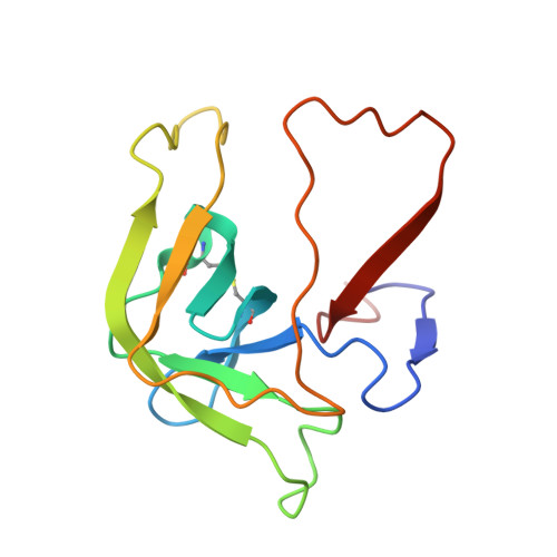
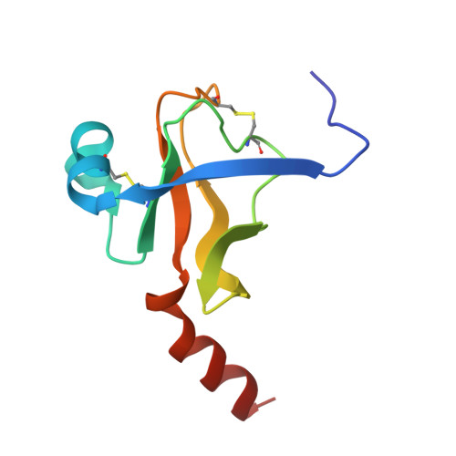
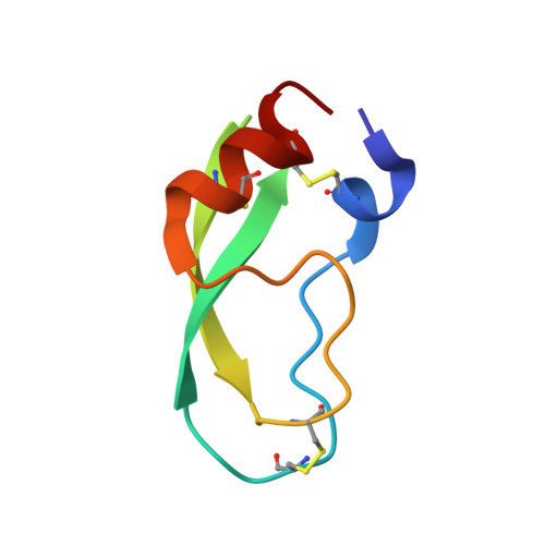
Fluorine-induced polarity increases inhibitory activity of BPTI towards chymotrypsin.

Leppkes, J.Dimos, N.Loll, B.Hohmann, T.Dyrks, M.Wieseke, A.Keller, B.G.Koksch, B.

(2022) RSC Chem Biol 3: 773-782

  • DOI: https://doi.org/10.1039/d2cb00018k
  • Primary Citation of Related Structures:  
    7QIQ, 7QIR, 7QIS, 7QIT

  • PubMed Abstract: 

    Substituting the P 1 position in bovine pancreatic trypsin inhibitor (BPTI) is known to heavily influence its inhibitory activity towards serine proteases. Side-chain fluorinated aliphatic amino acids have been shown to alter numerous properties of peptides and proteins and thus are of interest in the context of BPTI. In our study, we systematically investigated the site-specific incorporation of non-canonical amino acids into BPTI by microwave-assisted solid-phase peptide synthesis (SPPS). Inhibitor activity of the variants was tested towards the serine protease α-chymotrypsin. We observed enhanced inhibition of two fluorinated BPTIs compared to wild type and hydrocarbon variants. To further investigate the complexes, we performed X-ray structure analysis. Our findings underline the power fluorine offers as a tool in protein engineering to beneficially alter the effects on phenomena as protein-protein interactions.


  • Organizational Affiliation

    Department of Biology, Chemistry and Pharmacy, Institute of Chemistry and Biochemistry, Freie Universität Berlin Arnimallee 20 14195 Berlin Germany beate.koksch@fu-berlin.de.


Macromolecules

Find similar proteins by:  Sequence   |   3D Structure  

Entity ID: 1
MoleculeChains Sequence LengthOrganismDetailsImage
Chymotrypsin A chain A
A, E
13Bos taurusMutation(s): 0 
UniProt
Find proteins for P00766 (Bos taurus)
Explore P00766 
Go to UniProtKB:  P00766
Entity Groups  
Sequence Clusters30% Identity50% Identity70% Identity90% Identity95% Identity100% Identity
UniProt GroupP00766
Sequence Annotations
Expand
  • Reference Sequence
Find similar proteins by:  (by identity cutoff)  |  3D Structure
Entity ID: 2
MoleculeChains Sequence LengthOrganismDetailsImage
Chymotrypsin A chain B
B, F
131Bos taurusMutation(s): 0 
UniProt
Find proteins for P00766 (Bos taurus)
Explore P00766 
Go to UniProtKB:  P00766
Entity Groups  
Sequence Clusters30% Identity50% Identity70% Identity90% Identity95% Identity100% Identity
UniProt GroupP00766
Sequence Annotations
Expand
  • Reference Sequence
Find similar proteins by:  (by identity cutoff)  |  3D Structure
Entity ID: 3
MoleculeChains Sequence LengthOrganismDetailsImage
Chymotrypsin A chain C
C, G
97Bos taurusMutation(s): 0 
UniProt
Find proteins for P00766 (Bos taurus)
Explore P00766 
Go to UniProtKB:  P00766
Entity Groups  
Sequence Clusters30% Identity50% Identity70% Identity90% Identity95% Identity100% Identity
UniProt GroupP00766
Sequence Annotations
Expand
  • Reference Sequence
Find similar proteins by:  (by identity cutoff)  |  3D Structure
Entity ID: 4
MoleculeChains Sequence LengthOrganismDetailsImage
Pancreatic trypsin inhibitor
D, H
58Bos taurusMutation(s): 1 
UniProt
Find proteins for P00974 (Bos taurus)
Explore P00974 
Go to UniProtKB:  P00974
Entity Groups  
Sequence Clusters30% Identity50% Identity70% Identity90% Identity95% Identity100% Identity
UniProt GroupP00974
Sequence Annotations
Expand
  • Reference Sequence
Small Molecules
Ligands 2 Unique
IDChains Name / Formula / InChI Key2D Diagram3D Interactions
SO4
Query on SO4

Download Ideal Coordinates CCD File 
AA [auth C]
BA [auth C]
CA [auth C]
HA [auth D]
IA [auth D]
AA [auth C],
BA [auth C],
CA [auth C],
HA [auth D],
IA [auth D],
JA [auth D],
KA [auth D],
LA [auth D],
N [auth B],
O [auth B],
QA [auth F],
RA [auth F],
WA [auth G],
XA [auth G],
ZA [auth H]
SULFATE ION
O4 S
QAOWNCQODCNURD-UHFFFAOYSA-L
GOL
Query on GOL

Download Ideal Coordinates CCD File 
DA [auth D]
EA [auth D]
FA [auth D]
GA [auth D]
I [auth B]
DA [auth D],
EA [auth D],
FA [auth D],
GA [auth D],
I [auth B],
J [auth B],
K [auth B],
L [auth B],
M [auth B],
MA [auth F],
NA [auth F],
OA [auth F],
P [auth C],
PA [auth F],
Q [auth C],
R [auth C],
S [auth C],
SA [auth G],
T [auth C],
TA [auth G],
U [auth C],
UA [auth G],
V [auth C],
VA [auth G],
W [auth C],
X [auth C],
Y [auth C],
YA [auth H],
Z [auth C]
GLYCEROL
C3 H8 O3
PEDCQBHIVMGVHV-UHFFFAOYSA-N
Experimental Data & Validation

Experimental Data

  • Method: X-RAY DIFFRACTION
  • Resolution: 1.83 Å
  • R-Value Free: 0.189 
  • R-Value Work: 0.163 
  • R-Value Observed: 0.163 
  • Space Group: P 61
  • Diffraction Data: https://doi.org/10.18430/M37QIS
Unit Cell:
Length ( Å )Angle ( ˚ )
a = 100.269α = 90
b = 100.269β = 90
c = 206.215γ = 120
Software Package:
Software NamePurpose
PHENIXrefinement
XDSdata reduction
XSCALEdata scaling
PHASERphasing

Structure Validation

View Full Validation Report


Entry History & Funding Information

Deposition Data


Funding OrganizationLocationGrant Number
German Research Foundation (DFG)GermanyProject-ID 387284271

Revision History  (Full details and data files)

  • Version 1.0: 2022-03-09
    Type: Initial release
  • Version 1.1: 2022-06-01
    Changes: Database references
  • Version 1.2: 2022-07-06
    Changes: Database references
  • Version 1.3: 2024-01-31
    Changes: Data collection, Refinement description