7QIN | pdb_00007qin

In situ structure of actomyosin complex in skeletal sarcomere


Experimental Data Snapshot

  • Method: ELECTRON MICROSCOPY
  • Resolution: 6.60 Å
  • Aggregation State: TISSUE 
  • Reconstruction Method: SUBTOMOGRAM AVERAGING 

Starting Models: experimental
View more details

wwPDB Validation 3D Report Full Report

Validation slider image for 7QIN

This is version 1.2 of the entry. See complete history

Literature

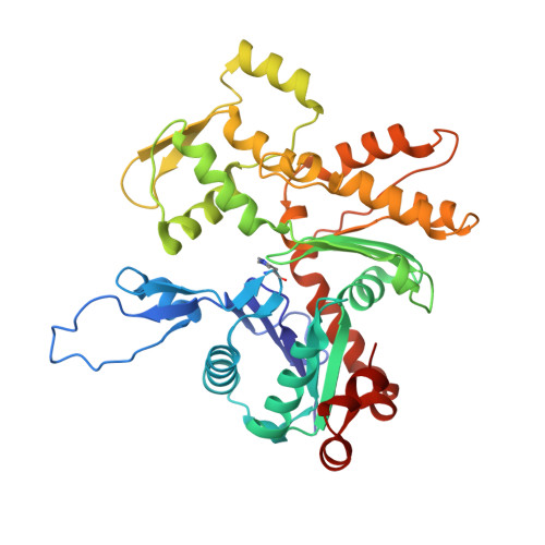
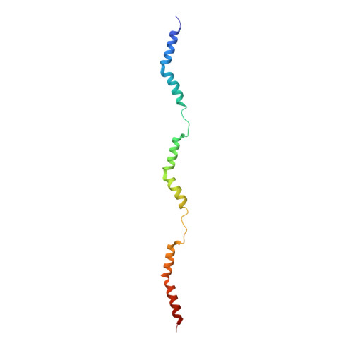
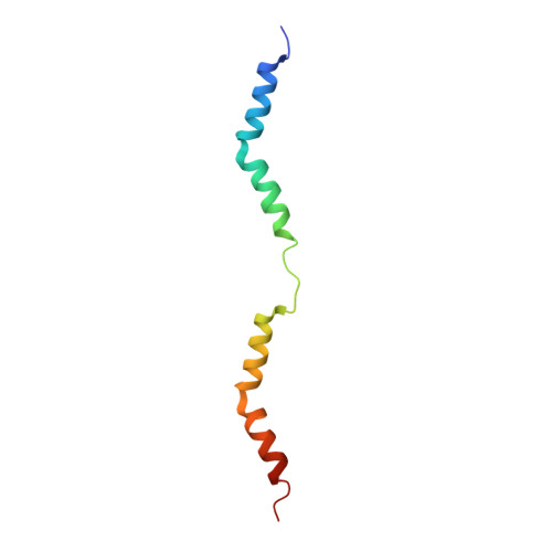
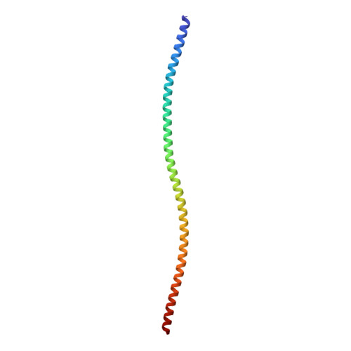
Structures from intact myofibrils reveal mechanism of thin filament regulation through nebulin.

Wang, Z.Grange, M.Pospich, S.Wagner, T.Kho, A.L.Gautel, M.Raunser, S.

(2022) Science 375: eabn1934-eabn1934

  • DOI: https://doi.org/10.1126/science.abn1934
  • Primary Citation Related Structures: 
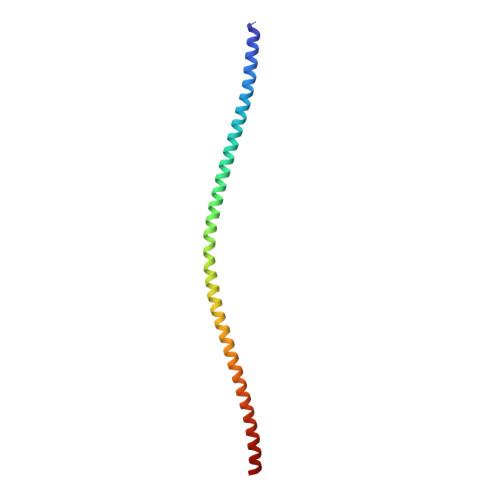
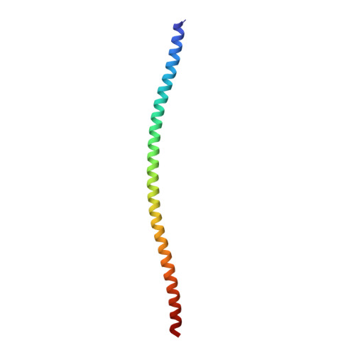
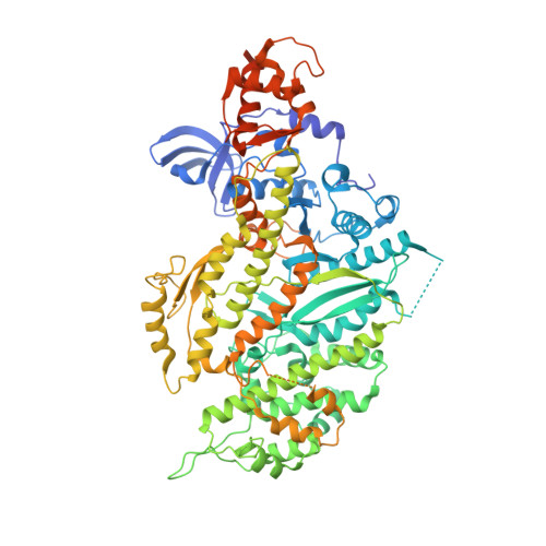
    7QIM, 7QIN, 7QIO

  • PubMed Abstract: 

    In skeletal muscle, nebulin stabilizes and regulates the length of thin filaments, but the underlying mechanism remains nebulous. In this work, we used cryo-electron tomography and subtomogram averaging to reveal structures of native nebulin bound to thin filaments within intact sarcomeres. This in situ reconstruction provided high-resolution details of the interaction between nebulin and actin, demonstrating the stabilizing role of nebulin. Myosin bound to the thin filaments exhibited different conformations of the neck domain, highlighting its inherent structural variability in muscle. Unexpectedly, nebulin did not interact with myosin or tropomyosin, but it did interact with a troponin T linker through two potential binding motifs on nebulin, explaining its regulatory role. Our structures support the role of nebulin as a thin filament "molecular ruler" and provide a molecular basis for studying nemaline myopathies.


  • Organizational Affiliation
    • Department of Structural Biochemistry, Max Planck Institute of Molecular Physiology, 44227 Dortmund, Germany.

Macromolecule Content 

  • Total Structure Weight: 452.37 kDa 
  • Atom Count: 29,589 
  • Modeled Residue Count: 3,939 
  • Deposited Residue Count: 4,137 
  • Unique protein chains: 7

Macromolecules

Find similar proteins by:|  3D Structure
Entity ID: 1
MoleculeChains  Sequence LengthOrganismDetailsImage
Actin, alpha skeletal muscle
A, B, C, D, E
371Mus musculusMutation(s): 0 
EC: 3.6.4
UniProt
Find proteins for P68134 (Mus musculus)
Explore P68134 
Go to UniProtKB:  P68134
Entity Groups
Sequence Clusters30% Identity50% Identity70% Identity90% Identity95% Identity100% Identity
UniProt GroupP68134
Sequence Annotations
Expand
Reference Sequence
Find similar proteins by:|  3D Structure
Entity ID: 2
MoleculeChains  Sequence LengthOrganismDetailsImage
nebulin (mouse)105Mus musculusMutation(s): 0 
Find similar proteins by:|  3D Structure
Entity ID: 3
MoleculeChains  Sequence LengthOrganismDetailsImage
nebulin (mouse)70Mus musculusMutation(s): 0 
Find similar proteins by:|  3D Structure
Entity ID: 4
MoleculeChains  Sequence LengthOrganismDetailsImage
tropomyosin, alpha-1 (mouse)117Mus musculusMutation(s): 0 
Find similar proteins by:|  3D Structure
Entity ID: 5
MoleculeChains  Sequence LengthOrganismDetailsImage
tropomyosin, alpha-1 (mouse)118Mus musculusMutation(s): 0 
Find similar proteins by:|  3D Structure
Entity ID: 6
MoleculeChains  Sequence LengthOrganismDetailsImage
tropomyosin, alpha-1 (mouse)
J, K
88Mus musculusMutation(s): 0 
Find similar proteins by:|  3D Structure
Entity ID: 7
MoleculeChains  Sequence LengthOrganismDetailsImage
Myosin-4
L, M
848Mus musculusMutation(s): 0 
UniProt & NIH Common Fund Data Resources
Find proteins for Q5SX39 (Mus musculus)
Explore Q5SX39 
Go to UniProtKB:  Q5SX39
IMPC:  MGI:1339713
Entity Groups
Sequence Clusters30% Identity50% Identity70% Identity90% Identity95% Identity100% Identity
UniProt GroupQ5SX39
Sequence Annotations
Expand
Reference Sequence

Small Molecules

Ligands 2 Unique
IDChains Name / Formula / InChI Key2D Diagram3D Interactions
ADP

Query on ADP


Download:Ideal Coordinates CCD File
N [auth A],
P [auth B],
R [auth C],
T [auth D],
V [auth E]
ADENOSINE-5'-DIPHOSPHATE
C10 H15 N5 O10 P2
XTWYTFMLZFPYCI-KQYNXXCUSA-N
MG

Query on MG


Download:Ideal Coordinates CCD File
O [auth A],
Q [auth B],
S [auth C],
U [auth D],
W [auth E]
MAGNESIUM ION
Mg
JLVVSXFLKOJNIY-UHFFFAOYSA-N
Modified Residues  1 Unique
IDChains TypeFormula2D DiagramParent
HIC
Query on HIC
A, B, C, D, E
L-PEPTIDE LINKINGC7 H11 N3 O2HIS
UNK
Query on UNK
F
L-PEPTIDE LINKINGC4 H9 N O2

--

Experimental Data & Validation

Experimental Data

  • Method: ELECTRON MICROSCOPY
  • Resolution: 6.60 Å
  • Aggregation State: TISSUE 
  • Reconstruction Method: SUBTOMOGRAM AVERAGING 
EM Software:
TaskSoftware PackageVersion
MODEL REFINEMENTPHENIX
MODEL REFINEMENTISOLDE

Structure Validation

View Full Validation Report


Entry History 

& Funding Information

Deposition Data


Funding OrganizationLocationGrant Number
Max Planck SocietyEuropean Union--
Wellcome TrustEuropean Union201543/Z/16/Z
European Research Council (ERC)European Union856118
Medical Research Council (MRC, United Kingdom)European UnionMR/R003106/1
European Molecular Biology Organization (EMBO)European Union--

Revision History  (Full details and data files)

  • Version 1.0: 2022-02-16
    Type: Initial release
  • Version 1.1: 2022-03-02
    Changes: Database references
  • Version 1.2: 2026-03-04
    Changes: Data collection, Refinement description, Structure summary