7QGB

H. SAPIENS CK2 KINASE ALPHA SUBUNIT IN COMPLEX WITH THE ATP-COMPETITIVE INHIBITOR 5,6-DIBROMOBENZOTRIAZOLE AT PH 6.5


Experimental Data Snapshot

  • Method: X-RAY DIFFRACTION
  • Resolution: 2.58 Å
  • R-Value Free: 0.234 
  • R-Value Work: 0.180 
  • R-Value Observed: 0.182 

wwPDB Validation   3D Report Full Report


Ligand Structure Quality Assessment 


This is version 1.2 of the entry. See complete history


Literature

Competition between electrostatic interactions and halogen bonding in the protein-ligand system: structural and thermodynamic studies of 5,6-dibromobenzotriazole-hCK2 alpha complexes.

Winiewska-Szajewska, M.Czapinska, H.Kaus-Drobek, M.Fricke, A.Mieczkowska, K.Dadlez, M.Bochtler, M.Poznanski, J.

(2022) Sci Rep 12: 18964-18964

  • DOI: https://doi.org/10.1038/s41598-022-23611-0
  • Primary Citation of Related Structures:  
    7QGB, 7QGC, 7QGD, 7QGE

  • PubMed Abstract: 

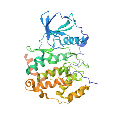
    CK2 is a member of the CMGC group of eukaryotic protein kinases and a cancer drug target. It can be efficiently inhibited by halogenated benzotriazoles and benzimidazoles. Depending on the scaffold, substitution pattern, and pH, these compounds are either neutral or anionic. Their binding poses are dictated by a hydrophobic effect (desolvation) and a tug of war between a salt bridge/hydrogen bond (to K68) and halogen bonding (to E114 and V116 backbone oxygens). Here, we test the idea that binding poses might be controllable by pH for ligands with near-neutral pK a , using the conditionally anionic 5,6-DBBt and constitutively anionic TBBt as our models. We characterize the binding by low-volume Differential Scanning Fluorimetry (nanoDSF), Isothermal Calorimetry (ITC), Hydrogen/Deuterium eXchange (HDX), and X-ray crystallography (MX). The data indicate that the ligand pose away from the hinge dominates for the entire tested pH range (5.5-8.5). The insensitivity of the binding mode to pH is attributed to the perturbation of ligand pK a upon binding that keeps it anionic in the ligand binding pocket at all tested pH values. However, a minor population of the ligand, detectable only by HDX, shifts towards the hinge in acidic conditions. Our findings demonstrate that electrostatic (ionic) interactions predominate over halogen bonding.


  • Organizational Affiliation

    Institute of Biochemistry and Biophysics PAS, Pawinskiego 5a, 02-106, Warsaw, Poland. mwin@ibb.waw.pl.


Macromolecules
Find similar proteins by:  (by identity cutoff)  |  3D Structure
Entity ID: 1
MoleculeChains Sequence LengthOrganismDetailsImage
Casein kinase II subunit alpha399Homo sapiensMutation(s): 0 
Gene Names: CSNK2A1CK2A1
EC: 2.7.11.1
UniProt & NIH Common Fund Data Resources
Find proteins for P68400 (Homo sapiens)
Explore P68400 
Go to UniProtKB:  P68400
PHAROS:  P68400
GTEx:  ENSG00000101266 
Entity Groups  
Sequence Clusters30% Identity50% Identity70% Identity90% Identity95% Identity100% Identity
UniProt GroupP68400
Sequence Annotations
Expand
  • Reference Sequence
Experimental Data & Validation

Experimental Data

  • Method: X-RAY DIFFRACTION
  • Resolution: 2.58 Å
  • R-Value Free: 0.234 
  • R-Value Work: 0.180 
  • R-Value Observed: 0.182 
  • Space Group: P 42 21 2
Unit Cell:
Length ( Å )Angle ( ˚ )
a = 128.487α = 90
b = 128.487β = 90
c = 61.239γ = 90
Software Package:
Software NamePurpose
XDSdata reduction
XDSdata scaling
MOLREPphasing
ARP/wARPmodel building
REFMACrefinement
PDB_EXTRACTdata extraction

Structure Validation

View Full Validation Report


Ligand Structure Quality Assessment 


Entry History & Funding Information

Deposition Data


Funding OrganizationLocationGrant Number
Polish National Science CentreEuropean Union2012/07/B/ST4/01334
Polish National Science CentreEuropean Union2017/25/B/ST4/01613
European Communitys Seventh Framework ProgrammeEuropean Union283570

Revision History  (Full details and data files)

  • Version 1.0: 2022-10-19
    Type: Initial release
  • Version 1.1: 2022-11-23
    Changes: Database references
  • Version 1.2: 2024-01-31
    Changes: Data collection, Refinement description