7QG7 | pdb_00007qg7

SARS-CoV-2 macrodomain Nsp3b bound to the remdesivir nucleoside GS-441524


Experimental Data Snapshot

  • Method: X-RAY DIFFRACTION
  • Resolution: 1.72 Å
  • R-Value Free: 
    0.226 (Depositor), 0.226 (DCC) 
  • R-Value Work: 
    0.183 (Depositor), 0.184 (DCC) 
  • R-Value Observed: 
    0.185 (Depositor) 

Starting Model: experimental
View more details

wwPDB Validation 3D Report Full Report

Validation slider image for 7QG7

Ligand Structure Quality Assessment 


This is version 1.3 of the entry. See complete history

Literature

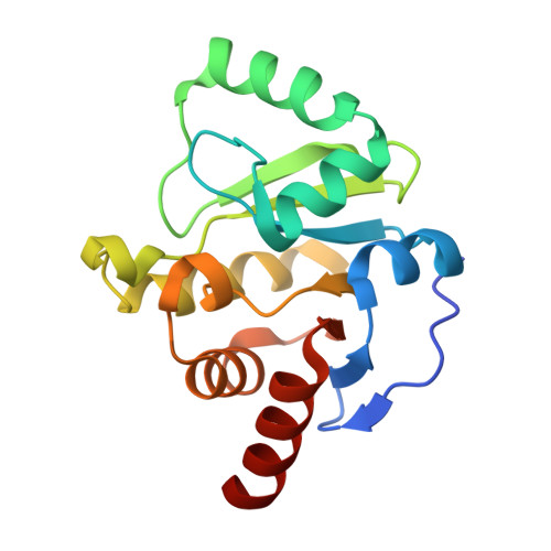
Binding Adaptation of GS-441524 Diversifies Macro Domains and Downregulates SARS-CoV-2 de-MARylation Capacity.

Tsika, A.C.Gallo, A.Fourkiotis, N.K.Argyriou, A.I.Sreeramulu, S.Lohr, F.Rogov, V.V.Richter, C.Linhard, V.Gande, S.L.Altincekic, N.Krishnathas, R.Elamri, I.Schwalbe, H.Wollenhaupt, J.Weiss, M.S.Spyroulias, G.A.

(2022) J Mol Biology 434: 167720-167720

  • DOI: https://doi.org/10.1016/j.jmb.2022.167720
  • Primary Citation Related Structures: 
    7P27, 7QG7

  • PubMed Abstract: 

    Viral infection in cells triggers a cascade of molecular defense mechanisms to maintain host-cell homoeostasis. One of these mechanisms is ADP-ribosylation, a fundamental post-translational modification (PTM) characterized by the addition of ADP-ribose (ADPr) on substrates. Poly(ADP-ribose) polymerases (PARPs) are implicated in this process and they perform ADP-ribosylation on host and pathogen proteins. Some viral families contain structural motifs that can reverse this PTM. These motifs known as macro domains (MDs) are evolutionarily conserved protein domains found in all kingdoms of life. They are divided in different classes with the viral belonging to Macro-D-type class because of their properties to recognize and revert the ADP-ribosylation. Viral MDs are potential pharmaceutical targets, capable to counteract host immune response. Sequence and structural homology between viral and human MDs are an impediment for the development of new active compounds against their function. Remdesivir, is a drug administrated in viral infections inhibiting viral replication through RNA-dependent RNA polymerase (RdRp). Herein, GS-441524, the active metabolite of the remdesivir, is tested as a hydrolase inhibitor for several viral MDs and for its binding to human homologs found in PARPs. This study presents biochemical and biophysical studies, which indicate that GS-441524 selectively modifies SARS-CoV-2 MD de-MARylation activity, while it does not interact with hPARP14 MD2 and hPARP15 MD2. The structural investigation of MD•GS-441524 complexes, using solution NMR and X-ray crystallography, discloses the impact of certain amino acids in ADPr binding cavity suggesting that F360 and its adjacent residues tune the selective binding of the inhibitor to SARS-CoV-2 MD.


  • Organizational Affiliation
    • Department of Pharmacy, University of Patras, GR-26504 Patras, Greece. Electronic address: https://twitter.com/@katerina_tsika.

Macromolecule Content 

  • Total Structure Weight: 38.25 kDa 
  • Atom Count: 2,889 
  • Modeled Residue Count: 339 
  • Deposited Residue Count: 346 
  • Unique protein chains: 1

Macromolecules

Find similar proteins by:|  3D Structure
Entity ID: 1
MoleculeChains  Sequence LengthOrganismDetailsImage
Papain-like protease nsp3
A, B
173Severe acute respiratory syndrome coronavirus 2Mutation(s): 0 
Gene Names: rep1a-1b
EC: 3.4.19.12 (PDB Primary Data), 3.4.22 (PDB Primary Data)
UniProt
Find proteins for P0DTD1 (Severe acute respiratory syndrome coronavirus 2)
Explore P0DTD1 
Go to UniProtKB:  P0DTD1
Entity Groups
Sequence Clusters30% Identity50% Identity70% Identity90% Identity95% Identity100% Identity
UniProt GroupP0DTD1
Sequence Annotations
Expand
Reference Sequence

Small Molecules

Ligands 2 Unique
IDChains Name / Formula / InChI Key2D Diagram3D Interactions
U08
(Subject of Investigation/LOI)

Query on U08


Download:Ideal Coordinates CCD File
C [auth A],
F [auth B]
(2~{R},3~{R},4~{S},5~{R})-2-(4-azanylpyrrolo[2,1-f][1,2,4]triazin-7-yl)-5-(hydroxymethyl)-3,4-bis(oxidanyl)oxolane-2-carbonitrile
C12 H13 N5 O4
BRDWIEOJOWJCLU-LTGWCKQJSA-N
EDO

Query on EDO


Download:Ideal Coordinates CCD File
D [auth A],
E [auth A],
G [auth B],
H [auth B],
I [auth B]
1,2-ETHANEDIOL
C2 H6 O2
LYCAIKOWRPUZTN-UHFFFAOYSA-N

Experimental Data & Validation

Experimental Data

  • Method: X-RAY DIFFRACTION
  • Resolution: 1.72 Å
  • R-Value Free:  0.226 (Depositor), 0.226 (DCC) 
  • R-Value Work:  0.183 (Depositor), 0.184 (DCC) 
  • R-Value Observed: 0.185 (Depositor) 
Space Group: P 2 21 21
Unit Cell:
Length ( Å )Angle ( ˚ )
a = 29.84α = 90
b = 73.87β = 90
c = 135.62γ = 90
Software Package:
Software NamePurpose
PHENIXrefinement
PHENIXrefinement
XDSdata reduction
XDSdata scaling
PHASERphasing

Structure Validation

View Full Validation Report


Ligand Structure Quality Assessment 


Entry History 

& Funding Information

Deposition Data


Funding OrganizationLocationGrant Number
European CommissionEuropean Union871037
European Union (EU)European Union20007375

Revision History  (Full details and data files)

  • Version 1.0: 2021-12-15
    Type: Initial release
  • Version 1.1: 2022-07-27
    Changes: Database references, Source and taxonomy, Structure summary
  • Version 1.2: 2022-08-03
    Changes: Database references
  • Version 1.3: 2024-01-31
    Changes: Data collection, Refinement description