The molecular architecture of Lactobacillus S-layer: Assembly and attachment to teichoic acids.
Sagmeister, T., Gubensak, N., Buhlheller, C., Grininger, C., Eder, M., Ðordic, A., Millan, C., Medina, A., Murcia, P.A.S., Berni, F., Hynonen, U., Vejzovic, D., Damisch, E., Kulminskaya, N., Petrowitsch, L., Oberer, M., Palva, A., Malanovic, N., Codee, J., Keller, W., Uson, I., Pavkov-Keller, T.(2024) Proc Natl Acad Sci U S A 121: e2401686121-e2401686121
- PubMed: 38838019 Search on PubMed
- DOI: https://doi.org/10.1073/pnas.2401686121
- Primary Citation Related Structures:
7QEC, 7QEH, 7QFG, 7QFI, 7QFJ, 7QFK, 7QFL, 7QLD, 7QLE, 7QLH, 8ALU, 8AOL, 8BT9, 8Q1O - PubMed Abstract:
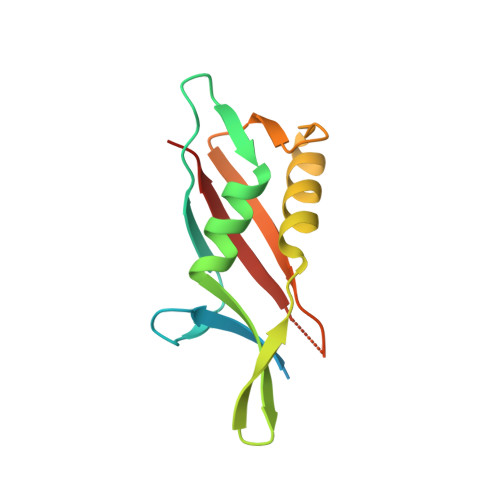
S-layers are crystalline arrays found on bacterial and archaeal cells. Lactobacillus is a diverse family of bacteria known especially for potential gut health benefits. This study focuses on the S-layer proteins from Lactobacillus acidophilus and Lactobacillus amylovorus common in the mammalian gut. Atomic resolution structures of Lactobacillus S-layer proteins SlpA and SlpX exhibit domain swapping, and the obtained assembly model of the main S-layer protein SlpA aligns well with prior electron microscopy and mutagenesis data. The S-layer's pore size suggests a protective role, with charged areas aiding adhesion. A highly similar domain organization and interaction network are observed across the Lactobacillus genus. Interaction studies revealed conserved binding areas specific for attachment to teichoic acids. The structure of the SlpA S-layer and the suggested incorporation of SlpX as well as its interaction with teichoic acids lay the foundation for deciphering its role in immune responses and for developing effective treatments for a variety of infectious and bacteria-mediated inflammation processes, opening opportunities for targeted engineering of the S-layer or lactobacilli bacteria in general.
- Institute of Molecular Biosciences, University of Graz, Graz, Austria 8010.
Organizational Affiliation: