7QF7

Orthorhombic crystal structure of PTG CBM21 in complex with beta-cyclodextrin


Experimental Data Snapshot

  • Method: X-RAY DIFFRACTION
  • Resolution: 1.47 Å
  • R-Value Free: 0.195 
  • R-Value Work: 0.175 
  • R-Value Observed: 0.176 

wwPDB Validation   3D Report Full Report


This is version 1.1 of the entry. See complete history


Literature

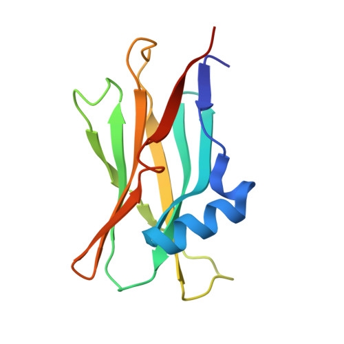
Molecular architecture of the glycogen- committed PP1/PTG holoenzyme.

Semrau, M.S.Giachin, G.Covaceuszach, S.Cassetta, A.Demitri, N.Storici, P.Lolli, G.

(2022) Nat Commun 13: 6199-6199

  • DOI: https://doi.org/10.1038/s41467-022-33693-z
  • Primary Citation of Related Structures:  
    7QF7, 7QFA, 7QFB, 7QM2

  • PubMed Abstract: 

    The delicate alternation between glycogen synthesis and degradation is governed by the interplay between key regulatory enzymes altering the activity of glycogen synthase and phosphorylase. Among these, the PP1 phosphatase promotes glycogenesis while inhibiting glycogenolysis. PP1 is, however, a master regulator of a variety of cellular processes, being conveniently directed to each of them by scaffolding subunits. PTG, Protein Targeting to Glycogen, addresses PP1 action to glycogen granules. In Lafora disease, the most aggressive pediatric epilepsy, genetic alterations leading to PTG accumulation cause the deposition of insoluble polyglucosans in neurons. Here, we report the crystallographic structure of the ternary complex PP1/PTG/carbohydrate. We further refine the mechanism of the PTG-mediated PP1 recruitment to glycogen by identifying i) an unusual combination of recruitment sites, ii) their contributions to the overall binding affinity, and iii) the conformational heterogeneity of this complex by in solution SAXS analyses.


  • Organizational Affiliation

    Department of Cellular, Computational and Integrative Biology - CIBio, University of Trento, via Sommarive 9, 38123, Povo, Trento, Italy.


Macromolecules
Find similar proteins by:  (by identity cutoff)  |  3D Structure
Entity ID: 1
MoleculeChains Sequence LengthOrganismDetailsImage
Protein phosphatase 1 regulatory subunit 3C134Homo sapiensMutation(s): 0 
Gene Names: PPP1R3CPPP1R5
UniProt & NIH Common Fund Data Resources
Find proteins for Q9UQK1 (Homo sapiens)
Explore Q9UQK1 
Go to UniProtKB:  Q9UQK1
PHAROS:  Q9UQK1
GTEx:  ENSG00000119938 
Entity Groups  
Sequence Clusters30% Identity50% Identity70% Identity90% Identity95% Identity100% Identity
UniProt GroupQ9UQK1
Sequence Annotations
Expand
  • Reference Sequence
Oligosaccharides

Help

Entity ID: 2
MoleculeChains Length2D Diagram Glycosylation3D Interactions
Cycloheptakis-(1-4)-(alpha-D-glucopyranose)B [auth C]7N/A
Glycosylation Resources
GlyTouCan:  G01435GL
GlyCosmos:  G01435GL
Small Molecules
Ligands 1 Unique
IDChains Name / Formula / InChI Key2D Diagram3D Interactions
NA
Query on NA

Download Ideal Coordinates CCD File 
C [auth A]SODIUM ION
Na
FKNQFGJONOIPTF-UHFFFAOYSA-N
Biologically Interesting Molecules (External Reference) 1 Unique
Experimental Data & Validation

Experimental Data

  • Method: X-RAY DIFFRACTION
  • Resolution: 1.47 Å
  • R-Value Free: 0.195 
  • R-Value Work: 0.175 
  • R-Value Observed: 0.176 
  • Space Group: P 2 21 21
Unit Cell:
Length ( Å )Angle ( ˚ )
a = 38.593α = 90
b = 56.443β = 90
c = 70.553γ = 90
Software Package:
Software NamePurpose
PHENIXrefinement
XDSdata reduction
Aimlessdata scaling
PHASERphasing
PDB_EXTRACTdata extraction

Structure Validation

View Full Validation Report


Entry History & Funding Information

Deposition Data


Funding OrganizationLocationGrant Number
Not funded--

Revision History  (Full details and data files)

  • Version 1.0: 2022-11-02
    Type: Initial release
  • Version 1.1: 2024-01-31
    Changes: Data collection, Refinement description