7QCT | pdb_00007qct

PDZ2 of LNX2 with SARS-CoV-2_E PBM complex


Experimental Data Snapshot

  • Method: X-RAY DIFFRACTION
  • Resolution: 3.20 Å
  • R-Value Free: 
    0.239 (Depositor), 0.228 (DCC) 
  • R-Value Work: 
    0.228 (Depositor) 
  • R-Value Observed: 
    0.229 (Depositor) 

Starting Model: experimental
View more details

wwPDB Validation 3D Report Full Report

Validation slider image for 7QCT

This is version 1.1 of the entry. See complete history

Literature

Interactions of Severe Acute Respiratory Syndrome Coronavirus 2 Protein E With Cell Junctions and Polarity PSD-95/Dlg/ZO-1-Containing Proteins.

Zhu, Y.Alvarez, F.Wolff, N.Mechaly, A.Brule, S.Neitthoffer, B.Etienne-Manneville, S.Haouz, A.Boeda, B.Caillet-Saguy, C.

(2022) Front Microbiol 13: 829094-829094

  • DOI: https://doi.org/10.3389/fmicb.2022.829094
  • Primary Citation Related Structures: 
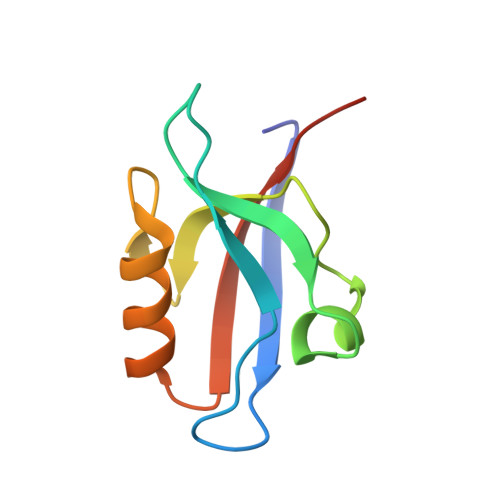
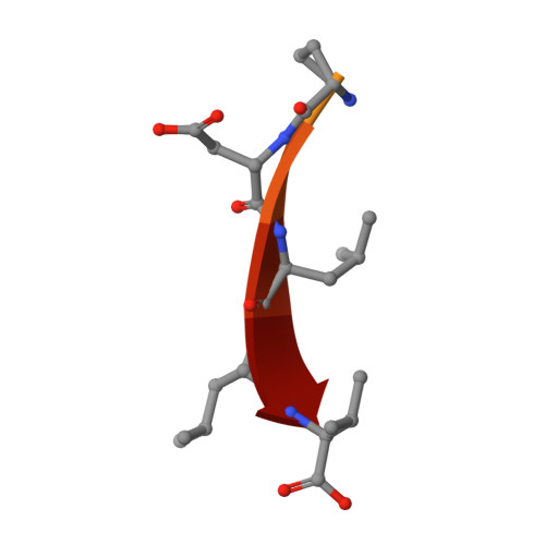
    7QCR, 7QCS, 7QCT

  • PubMed Abstract: 

    The C-terminus of the severe acute respiratory syndrome coronavirus 2 (SARS-CoV-2) protein E contains a PBM (PDZ-binding motif) targeting PDZ (PSD-95/Dlg/ZO-1) domains, which is identical to the PBM of SARS-CoV. The latter is involved in the pathogenicity of the virus. Recently, we identified 10 human PDZ-containing proteins showing significant interactions with SARS-CoV-2 protein E PBM. We selected several of them involved in cellular junctions and cell polarity (TJP1, PARD3, MLLT4, and LNX2) and MPP5/PALS1 previously shown to interact with SARS-CoV E PBM. Targeting cellular junctions and polarity components is a common strategy by viruses to hijack cell machinery to their advantage. In this study, we showed that these host PDZ domains TJP1, PARD3, MLLT4, LNX2, and MPP5/PALS1 interact in a PBM-dependent manner in vitro and colocalize with the full-length E protein in cellulo , sequestrating the PDZ domains to the Golgi compartment. We solved three crystal structures of complexes between human LNX2, MLLT4, and MPP5 PDZs and SARS-CoV-2 E PBM highlighting its binding preferences for several cellular targets. Finally, we showed different affinities for the PDZ domains with the original SARS-CoV-2 C-terminal sequence containing the PBM and the one of the beta variant that contains a mutation close to the PBM. The acquired mutations in the E protein localized near the PBM might have important effects both on the structure and the ion-channel activity of the E protein and on the host machinery targeted by the variants during the infection.


  • Organizational Affiliation
    • Channel Receptors Unit, CNRS, UMR 3571, Institut Pasteur, Université de Paris, Paris, France.

Macromolecule Content 

  • Total Structure Weight: 23.08 kDa 
  • Atom Count: 1,483 
  • Modeled Residue Count: 190 
  • Deposited Residue Count: 212 
  • Unique protein chains: 2

Macromolecules

Find similar proteins by:|  3D Structure
Entity ID: 1
MoleculeChains  Sequence LengthOrganismDetailsImage
Ligand of Numb protein X 2
A, B
94Homo sapiensMutation(s): 0 
Gene Names: LNX2PDZRN1
UniProt & NIH Common Fund Data Resources
Find proteins for Q8N448 (Homo sapiens)
Explore Q8N448 
Go to UniProtKB:  Q8N448
PHAROS:  Q8N448
GTEx:  ENSG00000139517 
Entity Groups
Sequence Clusters30% Identity50% Identity70% Identity90% Identity95% Identity100% Identity
UniProt GroupQ8N448
Sequence Annotations
Expand
Reference Sequence
Find similar proteins by:  Sequence   |   3D Structure  
Entity ID: 2
MoleculeChains  Sequence LengthOrganismDetailsImage
Envelope small membrane protein
C, D
12Severe acute respiratory syndrome coronavirus 2Mutation(s): 0 
UniProt
Find proteins for P0DTC4 (Severe acute respiratory syndrome coronavirus 2)
Explore P0DTC4 
Go to UniProtKB:  P0DTC4
Entity Groups
Sequence Clusters30% Identity50% Identity70% Identity90% Identity95% Identity100% Identity
UniProt GroupP0DTC4
Sequence Annotations
Expand
Reference Sequence

Experimental Data & Validation

Experimental Data

  • Method: X-RAY DIFFRACTION
  • Resolution: 3.20 Å
  • R-Value Free:  0.239 (Depositor), 0.228 (DCC) 
  • R-Value Work:  0.228 (Depositor) 
  • R-Value Observed: 0.229 (Depositor) 
Space Group: I 4 3 2
Unit Cell:
Length ( Å )Angle ( ˚ )
a = 183.23α = 90
b = 183.23β = 90
c = 183.23γ = 90
Software Package:
Software NamePurpose
BUSTERrefinement
XDSdata reduction
XDSdata scaling
PHASERphasing

Structure Validation

View Full Validation Report


Entry History 

& Funding Information

Deposition Data


Funding OrganizationLocationGrant Number
Pasteur InstituteFranceURGENCE COVID-19 fundraising campaign
Agence Nationale de la Recherche (ANR)FranceANR Recherche Action Covid19 FRM PDZCov2
Pasteur InstituteFranceDON MICHELIN COVID PFR-5 Cov-2-Cvnet

Revision History  (Full details and data files)

  • Version 1.0: 2022-04-20
    Type: Initial release
  • Version 1.1: 2024-01-31
    Changes: Data collection, Refinement description