7Q9N

Transthyretin complexed with (E)-4-(2-(naphthalen-2-yl)vinyl)benzene-1,2-diol


Experimental Data Snapshot

  • Method: X-RAY DIFFRACTION
  • Resolution: 1.45 Å
  • R-Value Free: 0.171 
  • R-Value Work: 0.146 
  • R-Value Observed: 0.147 

wwPDB Validation   3D Report Full Report


Ligand Structure Quality Assessment 


This is version 1.2 of the entry. See complete history


Literature

Transthyretin Binding Mode Dichotomy of Fluorescent trans -Stilbene Ligands.

Begum, A.Zhang, J.Derbyshire, D.Wu, X.Konradsson, P.Hammarstrom, P.von Castelmur, E.

(2023) ACS Chem Neurosci 14: 820-828

  • DOI: https://doi.org/10.1021/acschemneuro.2c00700
  • Primary Citation of Related Structures:  
    7Q9L, 7Q9N, 7Q9O, 8AWI

  • PubMed Abstract: 

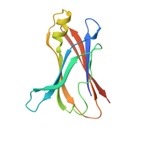
    The orientations of ligands bound to the transthyretin (TTR) thyroxine (T4) binding site are difficult to predict. Conflicting binding modes of resveratrol have been reported. We previously reported two resveratrol based trans -stilbene fluorescent ligands, ( E )-4-(2-(naphthalen-1-yl)vinyl)benzene-1,2-diol (SB-11) and ( E )-4-(2-(naphthalen-2-yl)vinyl)benzene-1,2-diol (SB-14), that bind native and misfolded protofibrillar TTR. The binding orientations of these two analogous ligands to native tetrameric TTR were predicted to be opposite. Herein we report the crystal structures of these TTR:ligand complexes. Opposite binding modes were verified but were different than predicted. The reverse binding mode (SB-14) placing the naphthalene moiety toward the opening of the binding pocket renders the fluorescent ligand pH sensitive due to changes in Lys15 amine protonation. Conversely, the forward binding mode (SB-11) placing the naphthalene inward mediates a stabilizing conformational change, allowing intersubunit H-bonding between Ser117 of different monomers across the dimer interface. Our structures of TTR complexes answer important questions in ligand design and interpretation of trans -stilbene binding modes to the TTR T4 binding site.


  • Organizational Affiliation

    Linköping University, IFM-Department of Physics, Chemistry and Biology, 58183 Linköping, Sweden.


Macromolecules
Find similar proteins by:  (by identity cutoff)  |  3D Structure
Entity ID: 1
MoleculeChains Sequence LengthOrganismDetailsImage
Transthyretin
A, B
119Homo sapiensMutation(s): 0 
Gene Names: TTRPALB
UniProt & NIH Common Fund Data Resources
Find proteins for P02766 (Homo sapiens)
Explore P02766 
Go to UniProtKB:  P02766
PHAROS:  P02766
GTEx:  ENSG00000118271 
Entity Groups  
Sequence Clusters30% Identity50% Identity70% Identity90% Identity95% Identity100% Identity
UniProt GroupP02766
Sequence Annotations
Expand
  • Reference Sequence
Small Molecules
Ligands 1 Unique
IDChains Name / Formula / InChI Key2D Diagram3D Interactions
9PS (Subject of Investigation/LOI)
Query on 9PS

Download Ideal Coordinates CCD File 
C [auth A],
D [auth B]
4-[(~{E})-2-naphthalen-2-ylethenyl]benzene-1,2-diol
C18 H14 O2
VBDICJKWECPUIF-AATRIKPKSA-N
Experimental Data & Validation

Experimental Data

  • Method: X-RAY DIFFRACTION
  • Resolution: 1.45 Å
  • R-Value Free: 0.171 
  • R-Value Work: 0.146 
  • R-Value Observed: 0.147 
  • Space Group: P 21 2 21
Unit Cell:
Length ( Å )Angle ( ˚ )
a = 42.545α = 90
b = 63.876β = 90
c = 85.137γ = 90
Software Package:
Software NamePurpose
XDSdata reduction
Aimlessdata scaling
PHASERphasing
REFMACrefinement
PDB_EXTRACTdata extraction

Structure Validation

View Full Validation Report


Ligand Structure Quality Assessment 


Entry History & Funding Information

Deposition Data


Funding OrganizationLocationGrant Number
Swedish Research CouncilSweden2019-04405

Revision History  (Full details and data files)

  • Version 1.0: 2022-11-23
    Type: Initial release
  • Version 1.1: 2023-03-01
    Changes: Database references
  • Version 1.2: 2023-03-15
    Changes: Database references