Vinylphosphonate-based cyclic dinucleotides enhance STING-mediated cancer immunotherapy.
Dejmek, M., Brazdova, A., Otava, T., Polidarova, M.P., Klima, M., Smola, M., Vavrina, Z., Budesinsky, M., Dracinsky, M., Liboska, R., Boura, E., Birkus, G., Nencka, R.(2023) Eur J Med Chem 259: 115685-115685
- PubMed: 37567057 Search on PubMed
- DOI: https://doi.org/10.1016/j.ejmech.2023.115685
- Primary Citation Related Structures:
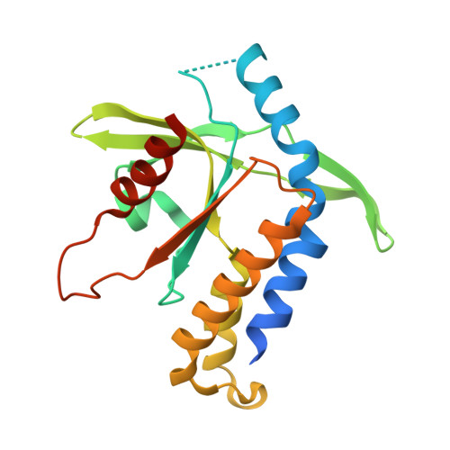
7Q85 - PubMed Abstract:
Cyclic dinucleotides (CDNs) trigger the cyclic GMP-AMP synthase-stimulator of interferon genes (cGAS-STING) pathway, which plays a key role in cytosolic DNA sensing and thus in immunomodulation against infections, cell damage and cancer. However, cancer immunotherapy trials with CDNs have shown immune activation, but not complete tumor regression. Nevertheless, we designed a novel class of CDNs containing vinylphosphonate based on a STING-affinity screening assay. In vitro, acyloxymethyl phosphate/phosphonate prodrugs of these vinylphosphonate CDNs were up to 1000-fold more potent than the clinical candidate ADU-S100. In vivo, the lead prodrug induced tumor-specific T cell priming and facilitated tumor regression in the 4T1 syngeneic mouse model of breast cancer. Moreover, we solved the crystal structure of this ligand bound to the STING protein. Therefore, our findings not only validate the therapeutic potential of vinylphosphonate CDNs but also open up opportunities for drug development in cancer immunotherapy bridging innate and adaptive immunity.
- Institute of Organic Chemistry and Biochemistry of the Czech Academy of Sciences, Flemingovo náměstí 2, Prague 6, 166 10, Czech Republic.
Organizational Affiliation: