7Q81

Crystal structure of the methyltransferase-ribozyme 1, 2'-Selenomethyl-Uridine modified (with 1-methyl-adenosine)


Experimental Data Snapshot

  • Method: X-RAY DIFFRACTION
  • Resolution: 2.85 Å
  • R-Value Free: 0.214 
  • R-Value Work: 0.188 
  • R-Value Observed: 0.189 

wwPDB Validation   3D Report Full Report


Ligand Structure Quality Assessment 


This is version 1.3 of the entry. See complete history


Literature

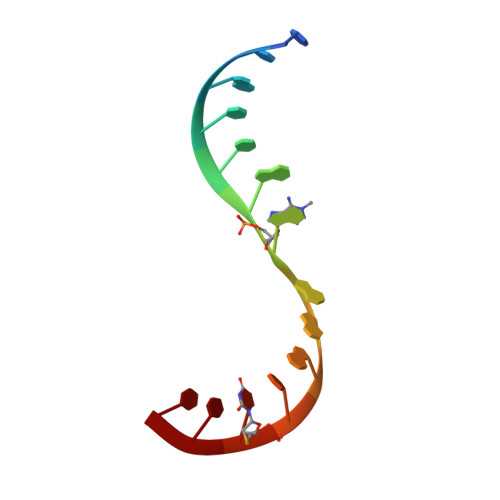
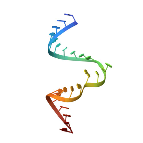
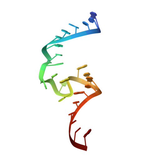
Structure and mechanism of the methyltransferase ribozyme MTR1.

Scheitl, C.P.M.Mieczkowski, M.Schindelin, H.Hobartner, C.

(2022) Nat Chem Biol 18: 547-555

  • DOI: https://doi.org/10.1038/s41589-022-00976-x
  • Primary Citation of Related Structures:  
    7Q7X, 7Q7Y, 7Q7Z, 7Q80, 7Q81, 7Q82

  • PubMed Abstract: 

    RNA-catalyzed RNA methylation was recently shown to be part of the catalytic repertoire of ribozymes. The methyltransferase ribozyme MTR1 catalyzes the site-specific synthesis of 1-methyladenosine (m 1 A) in RNA, using O 6 -methylguanine (m 6 G) as a methyl group donor. Here, we report the crystal structure of MTR1 at a resolution of 2.8 Å, which reveals a guanine-binding site reminiscent of natural guanine riboswitches. The structure represents the postcatalytic state of a split ribozyme in complex with the m 1 A-containing RNA product and the demethylated cofactor guanine. The structural data suggest the mechanistic involvement of a protonated cytidine in the methyl transfer reaction. A synergistic effect of two 2'-O-methylated ribose residues in the active site results in accelerated methyl group transfer. Supported by these results, it seems plausible that modified nucleotides may have enhanced early RNA catalysis and that metabolite-binding riboswitches may resemble inactivated ribozymes that have lost their catalytic activity during evolution.


  • Organizational Affiliation

    Institute of Organic Chemistry, Julius-Maximilians-Universität Würzburg, Am Hubland, Würzburg, Germany.


Macromolecules

Find similar nucleic acids by:  Sequence   |   3D Structure  

Entity ID: 1
MoleculeChains LengthOrganismImage
RNA114synthetic construct
Sequence Annotations
Expand
  • Reference Sequence

Find similar nucleic acids by:  Sequence   |   3D Structure  

Entity ID: 2
MoleculeChains LengthOrganismImage
RNA 224synthetic construct
Sequence Annotations
Expand
  • Reference Sequence

Find similar nucleic acids by:  Sequence   |   3D Structure  

Entity ID: 3
MoleculeChains LengthOrganismImage
RNA 324synthetic construct
Sequence Annotations
Expand
  • Reference Sequence
Experimental Data & Validation

Experimental Data

  • Method: X-RAY DIFFRACTION
  • Resolution: 2.85 Å
  • R-Value Free: 0.214 
  • R-Value Work: 0.188 
  • R-Value Observed: 0.189 
  • Space Group: P 41 21 2
Unit Cell:
Length ( Å )Angle ( ˚ )
a = 71.085α = 90
b = 71.085β = 90
c = 83.561γ = 90
Software Package:
Software NamePurpose
PHENIXrefinement
XDSdata reduction
Aimlessdata scaling
AutoSolphasing

Structure Validation

View Full Validation Report


Ligand Structure Quality Assessment 


Entry History & Funding Information

Deposition Data


Funding OrganizationLocationGrant Number
European Research Council (ERC)European Union--

Revision History  (Full details and data files)

  • Version 1.0: 2022-03-23
    Type: Initial release
  • Version 1.1: 2022-03-30
    Changes: Database references
  • Version 1.2: 2022-05-11
    Changes: Database references
  • Version 1.3: 2024-01-31
    Changes: Data collection, Refinement description