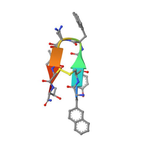
Atomic structure of Lanreotide nanotubes revealed by cryo-EM.
Pieri, L., Wang, F., Arteni, A.A., Vos, M., Winter, J.M., Le Du, M.H., Artzner, F., Gobeaux, F., Legrand, P., Boulard, Y., Bressanelli, S., Egelman, E.H., Paternostre, M.(2022) Proc Natl Acad Sci U S A 119
- PubMed: 35042822 Search on PubMedSearch on PubMed Central
- DOI: https://doi.org/10.1073/pnas.2120346119
- Primary Citation Related Structures:
7Q5A, 7Q5G - PubMed Abstract:
Functional and versatile nano- and microassemblies formed by biological molecules are found at all levels of life, from cell organelles to full organisms. Understanding the chemical and physicochemical determinants guiding the formation of these assemblies is crucial not only to understand the biological processes they carry out but also to mimic nature. Among the synthetic peptides forming well-defined nanostructures, the octapeptide Lanreotide has been considered one of the best characterized, in terms of both the atomic structure and its self-assembly process. In the present work, we determined the atomic structure of Lanreotide nanotubes at 2.5-Å resolution by cryoelectron microscopy (cryo-EM). Surprisingly, the asymmetric unit in the nanotube contains eight copies of the peptide, forming two tetramers. There are thus eight different environments for the peptide, and eight different conformations in the nanotube. The structure built from the cryo-EM map is strikingly different from the molecular model, largely based on X-ray fiber diffraction, proposed 20 y ago. Comparison of the nanotube with a crystal structure at 0.83-Å resolution of a Lanreotide derivative highlights the polymorphism for this peptide family. This work shows once again that higher-order assemblies formed by even well-characterized small peptides are very difficult to predict.
- CEA, CNRS, Université Paris-Saclay, Institute for Integrative Biology of the Cell (I2BC), 91198 Gif-sur-Yvette, France.
Organizational Affiliation: