


Binding of beta-lactam antibiotics by equine, caprine and ovine serum albumins
Sekula, B., Duszynski, K., Talaj, J., Bujacz, A.To be published.
Experimental Data Snapshot
Starting Model: experimental
View more details
Entity ID: 1 | |||||
|---|---|---|---|---|---|
| Molecule | Chains | Sequence Length | Organism | Details | Image |
| Albumin | 580 | Equus caballus | Mutation(s): 0 | 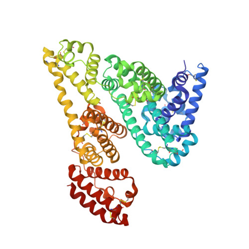 | |
UniProt | |||||
Entity Groups | |||||
| Sequence Clusters | 30% Identity50% Identity70% Identity90% Identity95% Identity100% Identity | ||||
| UniProt Group | P35747 | ||||
Sequence AnnotationsExpand | |||||
Reference Sequence | |||||
| Ligands 5 Unique | |||||
|---|---|---|---|---|---|
| ID | Chains | Name / Formula / InChI Key | 2D Diagram | 3D Interactions | |
| 8XI (Subject of Investigation/LOI) Download:Ideal Coordinates CCD File | B [auth A], C [auth A] | Cefaclor C15 H14 Cl N3 O4 S QYIYFLOTGYLRGG-GPCCPHFNSA-N |  | ||
| LMR Download:Ideal Coordinates CCD File | I [auth A] | (2S)-2-hydroxybutanedioic acid C4 H6 O5 BJEPYKJPYRNKOW-REOHCLBHSA-N |  | ||
| MLI Download:Ideal Coordinates CCD File | D [auth A], E [auth A], F [auth A], G [auth A], H [auth A] | MALONATE ION C3 H2 O4 OFOBLEOULBTSOW-UHFFFAOYSA-L |  | ||
| ACT Download:Ideal Coordinates CCD File | Q [auth A], R [auth A] | ACETATE ION C2 H3 O2 QTBSBXVTEAMEQO-UHFFFAOYSA-M |  | ||
| FMT Download:Ideal Coordinates CCD File | J [auth A] K [auth A] L [auth A] M [auth A] N [auth A] | FORMIC ACID C H2 O2 BDAGIHXWWSANSR-UHFFFAOYSA-N |  | ||
| Length ( Å ) | Angle ( ˚ ) |
|---|---|
| a = 94.75 | α = 90 |
| b = 94.75 | β = 90 |
| c = 142.62 | γ = 120 |
| Software Name | Purpose |
|---|---|
| REFMAC | refinement |
| PDB_EXTRACT | data extraction |
| XDS | data reduction |
| Coot | model building |
| XDS | data scaling |
| MOLREP | phasing |
| Funding Organization | Location | Grant Number |
|---|---|---|
| Polish National Science Centre | Poland | 2013/11/B/ST5/02271 |


