7Q4D

Local refinement structure of the two interacting N-domains of full-length, dimeric, soluble somatic angiotensin I-converting enzyme


Experimental Data Snapshot

  • Method: ELECTRON MICROSCOPY
  • Resolution: 3.78 Å
  • Aggregation State: PARTICLE 
  • Reconstruction Method: SINGLE PARTICLE 

wwPDB Validation   3D Report Full Report


This is version 1.2 of the entry. See complete history


Literature

Cryo-EM reveals mechanisms of angiotensin I-converting enzyme allostery and dimerization.

Lubbe, L.Sewell, B.T.Woodward, J.D.Sturrock, E.D.

(2022) EMBO J 41: e110550-e110550

  • DOI: https://doi.org/10.15252/embj.2021110550
  • Primary Citation of Related Structures:  
    7Q3Y, 7Q49, 7Q4C, 7Q4D, 7Q4E

  • PubMed Abstract: 

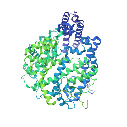
    Hypertension (high blood pressure) is a major risk factor for cardiovascular disease, which is the leading cause of death worldwide. The somatic isoform of angiotensin I-converting enzyme (sACE) plays a critical role in blood pressure regulation, and ACE inhibitors are thus widely used to treat hypertension and cardiovascular disease. Our current understanding of sACE structure, dynamics, function, and inhibition has been limited because truncated, minimally glycosylated forms of sACE are typically used for X-ray crystallography and molecular dynamics simulations. Here, we report the first cryo-EM structures of full-length, glycosylated, soluble sACE (sACE S1211 ). Both monomeric and dimeric forms of the highly flexible apo enzyme were reconstructed from a single dataset. The N- and C-terminal domains of monomeric sACE S1211 were resolved at 3.7 and 4.1 Å, respectively, while the interacting N-terminal domains responsible for dimer formation were resolved at 3.8 Å. Mechanisms are proposed for intradomain hinging, cooperativity, and homodimerization. Furthermore, the observation that both domains were in the open conformation has implications for the design of sACE modulators.


  • Organizational Affiliation

    Department of Integrative Biomedical Sciences, Institute of Infectious Disease and Molecular Medicine, University of Cape Town, Cape Town, South Africa.


Macromolecules
Find similar proteins by:  (by identity cutoff)  |  3D Structure
Entity ID: 1
MoleculeChains Sequence LengthOrganismDetailsImage
Angiotensin-converting enzyme
A, B
1,211Homo sapiensMutation(s): 1 
Gene Names: ACEDCPDCP1
EC: 3.2.1 (PDB Primary Data), 3.4.15.1 (PDB Primary Data)
UniProt & NIH Common Fund Data Resources
Find proteins for P12821 (Homo sapiens)
Explore P12821 
Go to UniProtKB:  P12821
PHAROS:  P12821
GTEx:  ENSG00000159640 
Entity Groups  
Sequence Clusters30% Identity50% Identity70% Identity90% Identity95% Identity100% Identity
UniProt GroupP12821
Sequence Annotations
Expand
  • Reference Sequence
Oligosaccharides

Help

Entity ID: 2
MoleculeChains Length2D Diagram Glycosylation3D Interactions
2-acetamido-2-deoxy-beta-D-glucopyranose-(1-2)-alpha-D-mannopyranose-(1-6)-[alpha-D-mannopyranose-(1-3)]beta-D-mannopyranose-(1-4)-2-acetamido-2-deoxy-beta-D-glucopyranose-(1-4)-[alpha-L-fucopyranose-(1-6)]2-acetamido-2-deoxy-beta-D-glucopyranose
C, M
7N-Glycosylation
Glycosylation Resources
GlyTouCan:  G45889JQ
GlyCosmos:  G45889JQ
GlyGen:  G45889JQ
Entity ID: 3
MoleculeChains Length2D Diagram Glycosylation3D Interactions
beta-D-mannopyranose-(1-4)-2-acetamido-2-deoxy-beta-D-glucopyranose-(1-4)-2-acetamido-2-deoxy-beta-D-glucopyranose
D, G, H, J, N
D, G, H, J, N, P
3N-Glycosylation
Glycosylation Resources
GlyTouCan:  G15407YE
GlyCosmos:  G15407YE
GlyGen:  G15407YE
Entity ID: 4
MoleculeChains Length2D Diagram Glycosylation3D Interactions
alpha-D-mannopyranose-(1-3)-beta-D-mannopyranose-(1-4)-2-acetamido-2-deoxy-beta-D-glucopyranose-(1-4)-2-acetamido-2-deoxy-beta-D-glucopyranose
E, K
4N-Glycosylation
Glycosylation Resources
GlyTouCan:  G81315DD
GlyCosmos:  G81315DD
GlyGen:  G81315DD
Entity ID: 5
MoleculeChains Length2D Diagram Glycosylation3D Interactions
2-acetamido-2-deoxy-beta-D-glucopyranose-(1-2)-alpha-D-mannopyranose-(1-6)-beta-D-mannopyranose-(1-4)-2-acetamido-2-deoxy-beta-D-glucopyranose-(1-4)-2-acetamido-2-deoxy-beta-D-glucopyranose
F, L
5N-Glycosylation
Glycosylation Resources
GlyTouCan:  G12045WP
GlyCosmos:  G12045WP
Entity ID: 6
MoleculeChains Length2D Diagram Glycosylation3D Interactions
2-acetamido-2-deoxy-beta-D-glucopyranose-(1-4)-2-acetamido-2-deoxy-beta-D-glucopyranose
I, O
2N-Glycosylation
Glycosylation Resources
GlyTouCan:  G42666HT
GlyCosmos:  G42666HT
GlyGen:  G42666HT
Experimental Data & Validation

Experimental Data

  • Method: ELECTRON MICROSCOPY
  • Resolution: 3.78 Å
  • Aggregation State: PARTICLE 
  • Reconstruction Method: SINGLE PARTICLE 
EM Software:
TaskSoftware PackageVersion
MODEL REFINEMENTPHENIX1.19.2
RECONSTRUCTIONcryoSPARC3.2

Structure Validation

View Full Validation Report


Entry History & Funding Information

Deposition Data


Funding OrganizationLocationGrant Number
UK Research and Innovation (UKRI)United KingdomST/R002754/1

Revision History  (Full details and data files)

  • Version 1.0: 2022-07-20
    Type: Initial release
  • Version 1.1: 2022-07-27
    Changes: Database references
  • Version 1.2: 2022-08-24
    Changes: Database references