7Q3B

Crystal structure of human STING in complex with 3'3'-c-(2'F,2'dA-isonucA)MP


Experimental Data Snapshot

  • Method: X-RAY DIFFRACTION
  • Resolution: 2.56 Å
  • R-Value Free: 0.232 
  • R-Value Work: 0.191 
  • R-Value Observed: 0.193 

wwPDB Validation   3D Report Full Report


Ligand Structure Quality Assessment 


This is version 1.4 of the entry. See complete history


Literature

Discovery of isonucleotidic CDNs as potent STING agonists with immunomodulatory potential.

Dejmek, M.Sala, M.Brazdova, A.Vanekova, L.Smola, M.Klima, M.Brehova, P.Budesinsky, M.Dracinsky, M.Prochazkova, E.Zavrel, M.Simak, O.Pav, O.Boura, E.Birkus, G.Nencka, R.

(2022) Structure 30: 1146

  • DOI: https://doi.org/10.1016/j.str.2022.05.012
  • Primary Citation of Related Structures:  
    7Q3B

  • PubMed Abstract: 

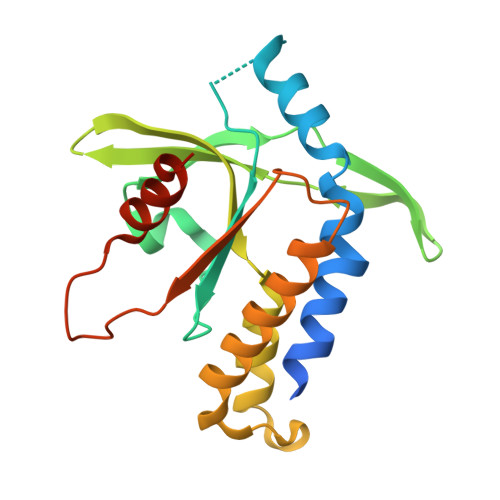
    Stimulator of interferon genes (STING) is an adaptor protein of the cGAS-STING signaling pathway involved in the sensing of cytosolic DNA. It functions as a receptor for cyclic dinucleotides (CDNs) and, upon their binding, mediates cytokine expression and host immunity. Besides naturally occurring CDNs, various synthetic CDNs, such as ADU-S100, have been reported to effectively activate STING and are being evaluated in clinical trials for the treatment of cancer. Here, we describe the preparation of a unique new class of STING agonists: isonucleotidic cyclic dinucleotides and the synthesis of their prodrugs. The presented CDNs stimulate STING with comparable efficiency to ADU-S100, whereas their prodrugs demonstrate activity up to four orders of magnitude better due to the improved cellular uptake. The compounds are very potent inducers of inflammatory cytokines by peripheral blood mononuclear cells (PBMCs). We also report the X-ray crystal structure of the lead inhibitor bound to the wild-type (WT) STING.


  • Organizational Affiliation

    Institute of Organic Chemistry and Biochemistry of the Czech Academy of Sciences, 16610 Prague, Czech Republic.


Macromolecules
Find similar proteins by:  (by identity cutoff)  |  3D Structure
Entity ID: 1
MoleculeChains Sequence LengthOrganismDetailsImage
Stimulator of interferon genes protein204Homo sapiensMutation(s): 1 
Gene Names: STING1ERISMITASTINGTMEM173
UniProt & NIH Common Fund Data Resources
Find proteins for Q86WV6 (Homo sapiens)
Explore Q86WV6 
Go to UniProtKB:  Q86WV6
PHAROS:  Q86WV6
GTEx:  ENSG00000184584 
Entity Groups  
Sequence Clusters30% Identity50% Identity70% Identity90% Identity95% Identity100% Identity
UniProt GroupQ86WV6
Sequence Annotations
Expand
  • Reference Sequence
Small Molecules
Ligands 1 Unique
IDChains Name / Formula / InChI Key2D Diagram3D Interactions
8SC (Subject of Investigation/LOI)
Query on 8SC

Download Ideal Coordinates CCD File 
B [auth A]3'3'-c-(2'F,2'dA-isonucA)MP
C20 H23 F N10 O10 P2
AGXHTOKVTXUJHH-HZQZQDQHSA-N
Experimental Data & Validation

Experimental Data

  • Method: X-RAY DIFFRACTION
  • Resolution: 2.56 Å
  • R-Value Free: 0.232 
  • R-Value Work: 0.191 
  • R-Value Observed: 0.193 
  • Space Group: P 41 21 2
Unit Cell:
Length ( Å )Angle ( ˚ )
a = 110.728α = 90
b = 110.728β = 90
c = 35.746γ = 90
Software Package:
Software NamePurpose
XDSdata reduction
XDSdata scaling
PHASERphasing
Cootmodel building
PHENIXrefinement

Structure Validation

View Full Validation Report


Ligand Structure Quality Assessment 


Entry History & Funding Information

Deposition Data


Funding OrganizationLocationGrant Number
Not funded--

Revision History  (Full details and data files)

  • Version 1.0: 2022-06-15
    Type: Initial release
  • Version 1.1: 2022-06-22
    Changes: Database references
  • Version 1.2: 2022-08-17
    Changes: Database references
  • Version 1.3: 2023-05-31
    Changes: Database references, Source and taxonomy, Structure summary
  • Version 1.4: 2024-02-07
    Changes: Data collection, Refinement description