7Q38 | pdb_00007q38

Crystal structure of the mutant bacteriorhodopsin pressurized with argon


Experimental Data Snapshot

  • Method: X-RAY DIFFRACTION
  • Resolution: 1.65 Å
  • R-Value Free: 
    0.205 (Depositor), 0.209 (DCC) 
  • R-Value Work: 
    0.162 (Depositor) 
  • R-Value Observed: 
    0.165 (Depositor) 

Starting Model: experimental
View more details

wwPDB Validation 3D Report Full Report

Validation slider image for 7Q38

This is version 1.2 of the entry. See complete history

Literature

High-pressure crystallography shows noble gas intervention into protein-lipid interaction and suggests a model for anaesthetic action.

Melnikov, I.Orekhov, P.Rulev, M.Kovalev, K.Astashkin, R.Bratanov, D.Ryzhykau, Y.Balandin, T.Bukhdruker, S.Okhrimenko, I.Borshchevskiy, V.Bourenkov, G.Mueller-Dieckmann, C.van der Linden, P.Carpentier, P.Leonard, G.Gordeliy, V.Popov, A.

(2022) Commun Biol 5: 360-360

  • DOI: https://doi.org/10.1038/s42003-022-03233-y
  • Primary Citation Related Structures: 
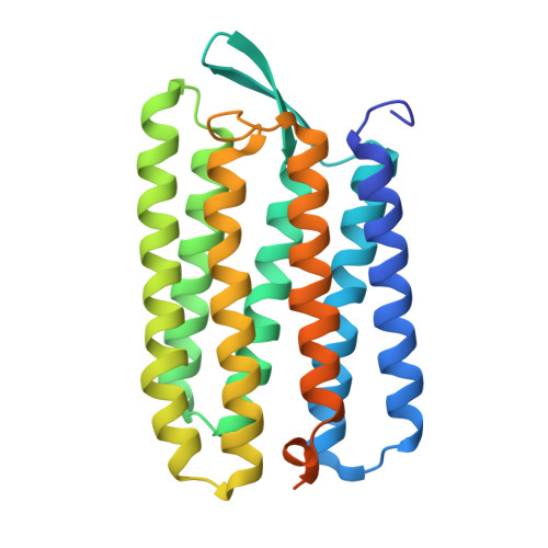
    7Q35, 7Q36, 7Q37, 7Q38

  • PubMed Abstract: 

    In this work we examine how small hydrophobic molecules such as inert gases interact with membrane proteins (MPs) at a molecular level. High pressure atmospheres of argon and krypton were used to produce noble gas derivatives of crystals of three well studied MPs (two different proton pumps and a sodium light-driven ion pump). The structures obtained using X-ray crystallography showed that the vast majority of argon and krypton binding sites were located on the outer hydrophobic surface of the MPs - a surface usually accommodating hydrophobic chains of annular lipids (which are known structural and functional determinants for MPs). In conformity with these results, supplementary in silico molecular dynamics (MD) analysis predicted even greater numbers of argon and krypton binding positions on MP surface within the bilayer. These results indicate a potential importance of such interactions, particularly as related to the phenomenon of noble gas-induced anaesthesia.


  • Organizational Affiliation
    • Institute of Biological Information Processing (IBI-7: Structural Biochemistry), Forschungszentrum Jülich GmbH, Jülich, Germany.

Macromolecule Content 

  • Total Structure Weight: 35.77 kDa 
  • Atom Count: 2,236 
  • Modeled Residue Count: 228 
  • Deposited Residue Count: 269 
  • Unique protein chains: 1

Macromolecules

Find similar proteins by:|  3D Structure
Entity ID: 1
MoleculeChains  Sequence LengthOrganismDetailsImage
Bacteriorhodopsin269Halobacterium salinarum NRC-1Mutation(s): 3 
Gene Names: bopVNG_1467G
Membrane Entity: Yes 
UniProt
Find proteins for P02945 (Halobacterium salinarum (strain ATCC 700922 / JCM 11081 / NRC-1))
Explore P02945 
Go to UniProtKB:  P02945
Entity Groups
Sequence Clusters30% Identity50% Identity70% Identity90% Identity95% Identity100% Identity
UniProt GroupP02945
Sequence Annotations
Expand
Reference Sequence

Small Molecules

Ligands 7 Unique
IDChains Name / Formula / InChI Key2D Diagram3D Interactions
OLC

Query on OLC


Download:Ideal Coordinates CCD File
M [auth A](2R)-2,3-dihydroxypropyl (9Z)-octadec-9-enoate
C21 H40 O4
RZRNAYUHWVFMIP-GDCKJWNLSA-N
RET

Query on RET


Download:Ideal Coordinates CCD File
B [auth A]RETINAL
C20 H28 O
NCYCYZXNIZJOKI-OVSJKPMPSA-N
LFA

Query on LFA


Download:Ideal Coordinates CCD File
C [auth A]
D [auth A]
E [auth A]
F [auth A]
G [auth A]
C [auth A],
D [auth A],
E [auth A],
F [auth A],
G [auth A],
H [auth A],
I [auth A],
J [auth A],
K [auth A],
L [auth A]
EICOSANE
C20 H42
CBFCDTFDPHXCNY-UHFFFAOYSA-N
OLA

Query on OLA


Download:Ideal Coordinates CCD File
N [auth A],
O [auth A],
P [auth A]
OLEIC ACID
C18 H34 O2
ZQPPMHVWECSIRJ-KTKRTIGZSA-N
SO4

Query on SO4


Download:Ideal Coordinates CCD File
U [auth A],
V [auth A]
SULFATE ION
O4 S
QAOWNCQODCNURD-UHFFFAOYSA-L
HEX

Query on HEX


Download:Ideal Coordinates CCD File
Q [auth A],
R [auth A],
S [auth A],
T [auth A]
HEXANE
C6 H14
VLKZOEOYAKHREP-UHFFFAOYSA-N
AR
(Subject of Investigation/LOI)

Query on AR


Download:Ideal Coordinates CCD File
AA [auth A]
AB [auth A]
BA [auth A]
BB [auth A]
CA [auth A]
AA [auth A],
AB [auth A],
BA [auth A],
BB [auth A],
CA [auth A],
CB [auth A],
DA [auth A],
DB [auth A],
EA [auth A],
EB [auth A],
FA [auth A],
FB [auth A],
GA [auth A],
GB [auth A],
HA [auth A],
HB [auth A],
IA [auth A],
IB [auth A],
JA [auth A],
JB [auth A],
KA [auth A],
KB [auth A],
LA [auth A],
LB [auth A],
MA [auth A],
MB [auth A],
NA [auth A],
NB [auth A],
OA [auth A],
OB [auth A],
PA [auth A],
PB [auth A],
QA [auth A],
QB [auth A],
RA [auth A],
SA [auth A],
TA [auth A],
UA [auth A],
VA [auth A],
W [auth A],
WA [auth A],
X [auth A],
XA [auth A],
Y [auth A],
YA [auth A],
Z [auth A],
ZA [auth A]
ARGON
Ar
XKRFYHLGVUSROY-UHFFFAOYSA-N

Experimental Data & Validation

Experimental Data

  • Method: X-RAY DIFFRACTION
  • Resolution: 1.65 Å
  • R-Value Free:  0.205 (Depositor), 0.209 (DCC) 
  • R-Value Work:  0.162 (Depositor) 
  • R-Value Observed: 0.165 (Depositor) 
Space Group: C 2 2 2
Unit Cell:
Length ( Å )Angle ( ˚ )
a = 115.657α = 90
b = 119.245β = 90
c = 36.393γ = 90
Software Package:
Software NamePurpose
REFMACrefinement
XDSdata reduction
Aimlessdata scaling
MOLREPphasing

Structure Validation

View Full Validation Report


Entry History 

& Funding Information

Deposition Data


Funding OrganizationLocationGrant Number
Not funded--

Revision History  (Full details and data files)

  • Version 1.0: 2022-04-27
    Type: Initial release
  • Version 1.1: 2024-01-31
    Changes: Data collection, Refinement description
  • Version 1.2: 2024-10-16
    Changes: Structure summary