Dynamic Long-Range Interactions Influence Substrate Binding and Catalysis by Human Histidine Triad Nucleotide-Binding Proteins (HINTs), Key Regulators of Multiple Cellular Processes and Activators of Antiviral ProTides.
Strom, A., Shah, R., Dolot, R., Rogers, M.S., Tong, C.L., Wang, D., Xia, Y., Lipscomb, J.D., Wagner, C.R.(2022) Biochemistry 61: 2648-2661
- PubMed: 36398895 Search on PubMedSearch on PubMed Central
- DOI: https://doi.org/10.1021/acs.biochem.2c00506
- Primary Citation Related Structures:
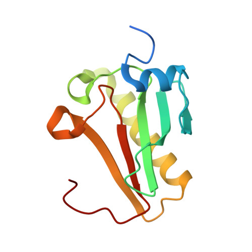
7Q2U - PubMed Abstract:
Human histidine triad nucleotide-binding (hHINT) proteins catalyze nucleotide phosphoramidase and acyl-phosphatase reactions that are essential for the activation of antiviral proTides, such as Sofosbuvir and Remdesivir. hHINT1 and hHINT2 are highly homologous but exhibit disparate roles as regulators of opioid tolerance (hHINT1) and mitochondrial activity (hHINT2). NMR studies of hHINT1 reveal a pair of dynamic surface residues (Q62, E100), which gate a conserved water channel leading to the active site 13 Å away. hHINT2 crystal structures identify analogous residues (R99, D137) and water channel. hHINT1 Q62 variants significantly alter the steady-state k cat and K m for turnover of the fluorescent substrate ( TpAd ), while stopped-flow kinetics indicate that K D also changes. hHINT2, like hHINT1, exhibits a burst phase of adenylation, monitored by fluorescent tryptamine release, prior to rate-limiting hydrolysis and nucleotide release. hHINT2 exhibits a much smaller burst-phase amplitude than hHINT1, which is further diminished in hHINT2 R99Q. Kinetic simulations suggest that amplitude variations can be accounted for by a variable fluorescent yield of the E·S complex from changes in the environment of bound TpAd . Isothermal titration calorimetry measurements of inhibitor binding show that these hHINT variants also alter the thermodynamic binding profile. We propose that these altered surface residues engender long-range dynamic changes that affect the orientation of bound ligands, altering the thermodynamic and kinetic characteristics of hHINT active site function. Thus, studies of the cellular roles and proTide activation potential by hHINTs should consider the importance of long-range interactions and possible protein binding surfaces far from the active site.
- Department of Medicinal Chemistry, University of Minnesota, Minneapolis, Minnesota55455, United States.
Organizational Affiliation: