7Q1U

Structure of Hedgehog acyltransferase (HHAT) in complex with megabody 177 bound to non-hydrolysable palmitoyl-CoA (Composite Map)


Experimental Data Snapshot

  • Method: ELECTRON MICROSCOPY
  • Resolution: 2.70 Å
  • Aggregation State: PARTICLE 
  • Reconstruction Method: SINGLE PARTICLE 

wwPDB Validation   3D Report Full Report


This is version 1.0 of the entry. See complete history


Literature

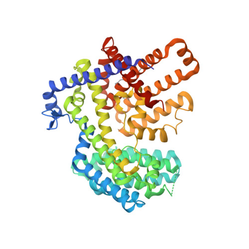
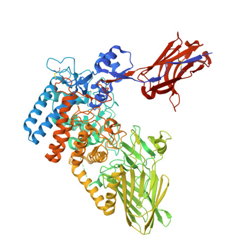
Structure, mechanism, and inhibition of Hedgehog acyltransferase.

Coupland, C.E.Andrei, S.A.Ansell, T.B.Carrique, L.Kumar, P.Sefer, L.Schwab, R.A.Byrne, E.F.X.Pardon, E.Steyaert, J.Magee, A.I.Lanyon-Hogg, T.Sansom, M.S.P.Tate, E.W.Siebold, C.

(2021) Mol Cell 81: 5025-5038.e10

  • DOI: https://doi.org/10.1016/j.molcel.2021.11.018
  • Primary Citation of Related Structures:  
    7Q1U, 7Q6Z

  • PubMed Abstract: 

    The Sonic Hedgehog (SHH) morphogen pathway is fundamental for embryonic development and stem cell maintenance and is implicated in various cancers. A key step in signaling is transfer of a palmitate group to the SHH N terminus, catalyzed by the multi-pass transmembrane enzyme Hedgehog acyltransferase (HHAT). We present the high-resolution cryo-EM structure of HHAT bound to substrate analog palmityl-coenzyme A and a SHH-mimetic megabody, revealing a heme group bound to HHAT that is essential for HHAT function. A structure of HHAT bound to potent small-molecule inhibitor IMP-1575 revealed conformational changes in the active site that occlude substrate binding. Our multidisciplinary analysis provides a detailed view of the mechanism by which HHAT adapts the membrane environment to transfer an acyl chain across the endoplasmic reticulum membrane. This structure of a membrane-bound O-acyltransferase (MBOAT) superfamily member provides a blueprint for other protein-substrate MBOATs and a template for future drug discovery.


  • Organizational Affiliation

    Division of Structural Biology, Wellcome Centre for Human Genetics, University of Oxford, Roosevelt Drive, Oxford OX3 7BN, UK.


Macromolecules
Find similar proteins by:  (by identity cutoff)  |  3D Structure
Entity ID: 1
MoleculeChains Sequence LengthOrganismDetailsImage
Protein-cysteine N-palmitoyltransferase HHAT503Homo sapiensMutation(s): 1 
Gene Names: HHATMART2SKI1
EC: 2.3.1
Membrane Entity: Yes 
UniProt & NIH Common Fund Data Resources
Find proteins for Q5VTY9 (Homo sapiens)
Explore Q5VTY9 
Go to UniProtKB:  Q5VTY9
PHAROS:  Q5VTY9
GTEx:  ENSG00000054392 
Entity Groups  
Sequence Clusters30% Identity50% Identity70% Identity90% Identity95% Identity100% Identity
UniProt GroupQ5VTY9
Sequence Annotations
Expand
  • Reference Sequence
Find similar proteins by:  (by identity cutoff)  |  3D Structure
Entity ID: 2
MoleculeChains Sequence LengthOrganismDetailsImage
Megabody 177919Escherichia coli K-12Mutation(s): 0 
Membrane Entity: Yes 
UniProt
Find proteins for P42592 (Escherichia coli (strain K12))
Explore P42592 
Go to UniProtKB:  P42592
Entity Groups  
Sequence Clusters30% Identity50% Identity70% Identity90% Identity95% Identity100% Identity
UniProt GroupP42592
Sequence Annotations
Expand
  • Reference Sequence
Small Molecules
Ligands 5 Unique
IDChains Name / Formula / InChI Key2D Diagram3D Interactions
HD6 (Subject of Investigation/LOI)
Query on HD6

Download Ideal Coordinates CCD File 
E [auth A][[(2~{R},3~{S},4~{R},5~{R})-5-(6-aminopurin-9-yl)-4-oxidanyl-3-phosphonooxy-oxolan-2-yl]methoxy-oxidanyl-phosphoryl] [(3~{S})-4-[[3-(2-hexadecylsulfanylethylamino)-3-oxidanylidene-propyl]amino]-2,2-dimethyl-3-oxidanyl-4-oxidanylidene-butyl] hydrogen phosphate
C37 H68 N7 O16 P3 S
HCCBNDDJPKNSLM-HNOUMJPASA-N
HEM (Subject of Investigation/LOI)
Query on HEM

Download Ideal Coordinates CCD File 
D [auth A]PROTOPORPHYRIN IX CONTAINING FE
C34 H32 Fe N4 O4
KABFMIBPWCXCRK-RGGAHWMASA-L
CLR
Query on CLR

Download Ideal Coordinates CCD File 
C [auth A]CHOLESTEROL
C27 H46 O
HVYWMOMLDIMFJA-DPAQBDIFSA-N
PLM
Query on PLM

Download Ideal Coordinates CCD File 
G [auth A],
H [auth A]
PALMITIC ACID
C16 H32 O2
IPCSVZSSVZVIGE-UHFFFAOYSA-N
MG
Query on MG

Download Ideal Coordinates CCD File 
F [auth A]MAGNESIUM ION
Mg
JLVVSXFLKOJNIY-UHFFFAOYSA-N
Modified Residues  1 Unique
IDChains TypeFormula2D DiagramParent
P1L
Query on P1L
A
L-PEPTIDE LINKINGC19 H37 N O3 SCYS
Experimental Data & Validation

Experimental Data

  • Method: ELECTRON MICROSCOPY
  • Resolution: 2.70 Å
  • Aggregation State: PARTICLE 
  • Reconstruction Method: SINGLE PARTICLE 
EM Software:
TaskSoftware PackageVersion
RECONSTRUCTIONcryoSPARCV3.1.1
MODEL REFINEMENTPHENIX1.19.2
MODEL REFINEMENTCoot0.9.2

Structure Validation

View Full Validation Report


Entry History & Funding Information

Deposition Data


Funding OrganizationLocationGrant Number
Cancer Research UKEuropean UnionC20724/A26752
Cancer Research UKEuropean UnionC20724/A14414
Biotechnology and Biological Sciences Research Council (BBSRC)European UnionBB/T01508X/1
European Research Council (ERC)European Union647278

Revision History  (Full details and data files)

  • Version 1.0: 2022-06-08
    Type: Initial release