Crystal structure and metal binding properties of the periplasmic iron component EfeM from Pseudomonas syringae EfeUOB/M iron-transport system.
Rajasekaran, M.B., Hussain, R., Siligardi, G., Andrews, S.C., Watson, K.A.(2022) Biometals 35: 573-589
- PubMed: 35348940 Search on PubMedSearch on PubMed Central
- DOI: https://doi.org/10.1007/s10534-022-00389-2
- Primary Citation Related Structures:
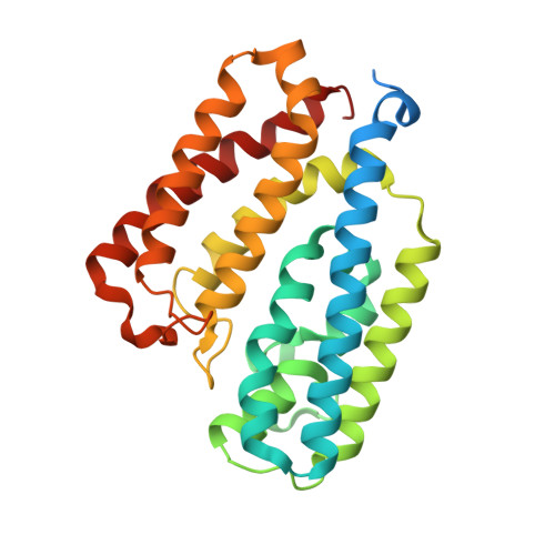
7Q1G - PubMed Abstract:
EfeUOB/M has been characterised in Pseudomonas syringae pathovar. syringae as a novel type of ferrous-iron transporter, consisting of an inner-membrane protein (EfeU Psy ) and three periplasmic proteins (EfeO Psy , EfeM Psy and EfeB Psy ). The role of an iron permease and peroxidase function has been identified for the EfeU and EfeB proteins, respectively, but the role of EfeO/M remains unclear. EfeM Psy is an 'M75-only' EfeO-like protein with a C-terminal peptidase-M75 domain (EfeO II /EfeM family). Herein, we report the 1.6 Å resolution crystal structure of EfeM Psy , the first structural report for an EfeM component of P. syringae pv. syringae. The structure possesses the bi-lobate architecture found in other bacterial periplasmic substrate/solute binding proteins. Metal binding studies, using SRCD and ICP-OES, reveal a preference of EfeM Psy for copper, iron and zinc. This work provides detailed knowledge of the structural scaffold, the metal site geometry, and the divalent metal binding potential of EfeM. This work provides crucial underpinning for a more detailed understanding of the role of EfeM/EfeO proteins and the peptidase-M75 domains in EfeUOB/M iron uptake systems in bacteria.
- School of Biological Sciences, Health and Life Sciences Building, University of Reading, Whiteknights Campus, Reading, RG6 6EX, UK.
Organizational Affiliation: