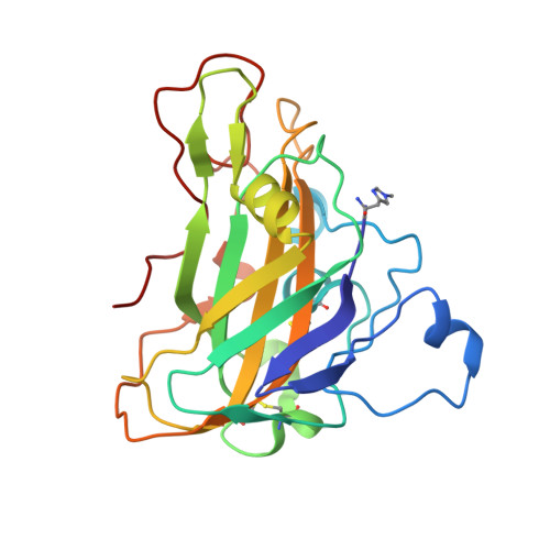
Changes in active-site geometry on X-ray photoreduction of a lytic polysaccharide monooxygenase active-site copper and saccharide binding.
Tandrup, T., Muderspach, S.J., Banerjee, S., Santoni, G., Ipsen, J.O., Hernandez-Rollan, C., Norholm, M.H.H., Johansen, K.S., Meilleur, F., Lo Leggio, L.(2022) IUCrJ 9: 666-681
- PubMed: 36071795 Search on PubMedSearch on PubMed Central
- DOI: https://doi.org/10.1107/S2052252522007175
- Primary Citation Related Structures:
7PQR, 7PXI, 7PXJ, 7PXK, 7PXL, 7PXM, 7PXN, 7PXR, 7PXS, 7PXT, 7PXU, 7PXV, 7PXW, 7PYD, 7PYE, 7PYF, 7PYG, 7PYH, 7PYI, 7PYL, 7PYM, 7PYN, 7PYO, 7PYP, 7PYQ, 7PYU, 7PYW, 7PYX, 7PYY, 7PYZ, 7PZ0, 7PZ3, 7PZ4, 7PZ5, 7PZ6, 7PZ7, 7PZ8 - PubMed Abstract:
The recently discovered lytic polysaccharide monooxygenases (LPMOs) are Cu-containing enzymes capable of degrading polysaccharide substrates oxidatively. The generally accepted first step in the LPMO reaction is the reduction of the active-site metal ion from Cu 2+ to Cu + . Here we have used a systematic diffraction data collection method to monitor structural changes in two AA9 LPMOs, one from Lentinus similis ( Ls AA9_A) and one from Thermoascus auranti-acus ( Ta AA9_A), as the active-site Cu is photoreduced in the X-ray beam. For Ls AA9_A, the protein produced in two different recombinant systems was crystallized to probe the effect of post-translational modifications and different crystallization conditions on the active site and metal photoreduction. We can recommend that crystallographic studies of AA9 LPMOs wishing to address the Cu 2+ form use a total X-ray dose below 3 × 10 4 Gy, while the Cu + form can be attained using 1 × 10 6 Gy. In all cases, we observe the transition from a hexa-coordinated Cu site with two solvent-facing ligands to a T-shaped geometry with no exogenous ligands, and a clear increase of the θ 2 parameter and a decrease of the θ 3 parameter by averages of 9.2° and 8.4°, respectively, but also a slight increase in θ T . Thus, the θ 2 and θ 3 parameters are helpful diagnostics for the oxidation state of the metal in a His-brace protein. On binding of cello-oligosaccharides to Ls AA9_A, regardless of the production source, the θ T parameter increases, making the Cu site less planar, while the active-site Tyr-Cu distance decreases reproducibly for the Cu 2+ form. Thus, the θ T increase found on copper reduction may bring Ls AA9_A closer to an oligosaccharide-bound state and contribute to the observed higher affinity of reduced Ls AA9_A for cellulosic substrates.
- Department of Chemistry, University of Copenhagen, Universitetsparken 5, 2100-DK, Copenhagen, Denmark.
Organizational Affiliation: