7PXX

The crystal structure of Leishmania major Pteridine Reductase 1 in complex with substrate folic acid


Experimental Data Snapshot

  • Method: X-RAY DIFFRACTION
  • Resolution: 1.81 Å
  • R-Value Free: 0.267 
  • R-Value Work: 0.227 
  • R-Value Observed: 0.229 

wwPDB Validation   3D Report Full Report


Ligand Structure Quality Assessment 


This is version 1.2 of the entry. See complete history


Literature

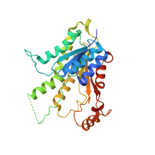
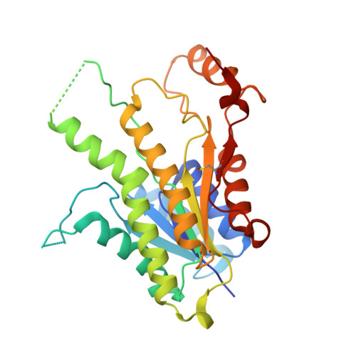
Crystal structure of the ternary complex of Leishmania major pteridine reductase 1 with the cofactor NADP + /NADPH and the substrate folic acid.

Dello Iacono, L.Di Pisa, F.Mangani, S.

(2022) Acta Crystallogr F Struct Biol Commun 78: 170-176

  • DOI: https://doi.org/10.1107/S2053230X22002795
  • Primary Citation of Related Structures:  
    7PXX

  • PubMed Abstract: 

    Pteridine reductase 1 (PTR1) is a key enzyme of the folate pathway in protozoan parasites of the genera Leishmania and Trypanosoma and is a valuable drug target for tropical diseases. This enzyme is able to catalyze the NADPH-dependent reduction of both conjugated (folate) and unconjugated (biopterin) pterins to their tetrahydro forms, starting from oxidized- or dihydro-state substrates. The currently available X-ray structures of Leishmania major PTR1 (LmPTR1) show the enzyme in its unbound, unconjugated substrate-bound (with biopterin derivatives) and inhibitor-bound forms. However, no structure has yet been determined of LmPTR1 bound to a conjugated substrate. Here, the high-resolution crystal structure of LmPTR1 in complex with folic acid is presented and the intermolecular forces that drive the binding of the substrate in the catalytic pocket are described. By expanding the collection of LmPTR1 structures in complex with process intermediates, additional insights into the active-site rearrangements that occur during the catalytic process are provided. In contrast to previous structures with biopterin derivatives, a small but significant difference in the orientation of Asp181 and Tyr194 of the catalytic triad is found. This feature is shared by PTR1 from T. brucei (TbPTR1) in complex with the same substrate molecule and may be informative in deciphering the importance of such residues at the beginning of the catalytic process.


  • Organizational Affiliation

    Department of Biotechnology, Chemistry and Pharmacy, University of Siena, Via Aldo Moro 2, 53100 Siena, Italy.


Macromolecules
Find similar proteins by:  (by identity cutoff)  |  3D Structure
Entity ID: 1
MoleculeChains Sequence LengthOrganismDetailsImage
Pteridine reductase 1288Leishmania majorMutation(s): 0 
Gene Names: PTR1HMTXRL1063.01LmjF23.0270LmjF_23_0270
EC: 1.5.1.33
UniProt
Find proteins for Q01782 (Leishmania major)
Explore Q01782 
Go to UniProtKB:  Q01782
Entity Groups  
Sequence Clusters30% Identity50% Identity70% Identity90% Identity95% Identity100% Identity
UniProt GroupQ01782
Sequence Annotations
Expand
  • Reference Sequence
Find similar proteins by:  (by identity cutoff)  |  3D Structure
Entity ID: 2
MoleculeChains Sequence LengthOrganismDetailsImage
Pteridine reductase 1
B, C, D
288Leishmania majorMutation(s): 0 
Gene Names: PTR1HMTXRL1063.01LmjF23.0270LmjF_23_0270
EC: 1.5.1.33
UniProt
Find proteins for Q01782 (Leishmania major)
Explore Q01782 
Go to UniProtKB:  Q01782
Entity Groups  
Sequence Clusters30% Identity50% Identity70% Identity90% Identity95% Identity100% Identity
UniProt GroupQ01782
Sequence Annotations
Expand
  • Reference Sequence
Small Molecules
Ligands 5 Unique
IDChains Name / Formula / InChI Key2D Diagram3D Interactions
NDP (Subject of Investigation/LOI)
Query on NDP

Download Ideal Coordinates CCD File 
F [auth A],
I [auth B],
P [auth C],
R [auth D]
NADPH DIHYDRO-NICOTINAMIDE-ADENINE-DINUCLEOTIDE PHOSPHATE
C21 H30 N7 O17 P3
ACFIXJIJDZMPPO-NNYOXOHSSA-N
FOL (Subject of Investigation/LOI)
Query on FOL

Download Ideal Coordinates CCD File 
E [auth A],
H [auth B],
O [auth C],
Q [auth D]
FOLIC ACID
C19 H19 N7 O6
OVBPIULPVIDEAO-LBPRGKRZSA-N
PGE
Query on PGE

Download Ideal Coordinates CCD File 
G [auth A],
J [auth B]
TRIETHYLENE GLYCOL
C6 H14 O4
ZIBGPFATKBEMQZ-UHFFFAOYSA-N
PEG
Query on PEG

Download Ideal Coordinates CCD File 
N [auth B]DI(HYDROXYETHYL)ETHER
C4 H10 O3
MTHSVFCYNBDYFN-UHFFFAOYSA-N
GOL
Query on GOL

Download Ideal Coordinates CCD File 
K [auth B],
L [auth B],
M [auth B],
S [auth D]
GLYCEROL
C3 H8 O3
PEDCQBHIVMGVHV-UHFFFAOYSA-N
Modified Residues  1 Unique
IDChains TypeFormula2D DiagramParent
CSX
Query on CSX
A
L-PEPTIDE LINKINGC3 H7 N O3 SCYS
Experimental Data & Validation

Experimental Data

  • Method: X-RAY DIFFRACTION
  • Resolution: 1.81 Å
  • R-Value Free: 0.267 
  • R-Value Work: 0.227 
  • R-Value Observed: 0.229 
  • Space Group: P 21 21 21
Unit Cell:
Length ( Å )Angle ( ˚ )
a = 94.9α = 90
b = 103.75β = 90
c = 136.79γ = 90
Software Package:
Software NamePurpose
PHENIXrefinement
MOSFLMdata reduction
SCALAdata scaling
MOLREPphasing

Structure Validation

View Full Validation Report


Ligand Structure Quality Assessment 


Entry History & Funding Information

Deposition Data


Funding OrganizationLocationGrant Number
European Union (EU)European Union603240

Revision History  (Full details and data files)

  • Version 1.0: 2022-04-06
    Type: Initial release
  • Version 1.1: 2022-04-20
    Changes: Database references
  • Version 1.2: 2024-01-31
    Changes: Data collection, Refinement description