


Crystal structure of the glutaredoxin/ferredoxin disulfide reductase fusion protein from Desulfotalea psychrophila Lsv54
Zannini, F., Mathiot, S., Couturier, J., Didierjean, C., Rouhier, N.(2022) Inorganics
Experimental Data Snapshot
wwPDB Validation 3D Report Full Report

(2022) Inorganics
Entity ID: 1 | |||||
|---|---|---|---|---|---|
| Molecule | Chains | Sequence Length | Organism | Details | Image |
| Ferredoxin-thioredoxin reductase subunit B | 196 | Desulfotalea psychrophila LSv54 | Mutation(s): 0 Gene Names: DP2155 EC: 1.8.7.2 | 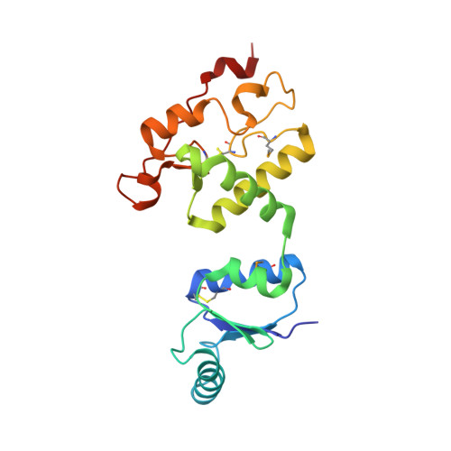 | |
UniProt | |||||
Entity Groups | |||||
| Sequence Clusters | 30% Identity50% Identity70% Identity90% Identity95% Identity100% Identity | ||||
| UniProt Group | Q6AL91 | ||||
Sequence AnnotationsExpand | |||||
Reference Sequence | |||||
| Ligands 2 Unique | |||||
|---|---|---|---|---|---|
| ID | Chains | Name / Formula / InChI Key | 2D Diagram | 3D Interactions | |
| SF4 (Subject of Investigation/LOI) Download:Ideal Coordinates CCD File | C [auth A], H [auth B] | IRON/SULFUR CLUSTER Fe4 S4 LJBDFODJNLIPKO-UHFFFAOYSA-N |  | ||
| IMD Download:Ideal Coordinates CCD File | D [auth A] E [auth A] F [auth A] G [auth A] I [auth B] | IMIDAZOLE C3 H5 N2 RAXXELZNTBOGNW-UHFFFAOYSA-O |  | ||
| Modified Residues 1 Unique | |||||
|---|---|---|---|---|---|
| ID | Chains | Type | Formula | 2D Diagram | Parent |
| MSE Query on MSE | A, B | L-PEPTIDE LINKING | C5 H11 N O2 Se |  | MET |
| Length ( Å ) | Angle ( ˚ ) |
|---|---|
| a = 123.6 | α = 90 |
| b = 49.3 | β = 116.5 |
| c = 86 | γ = 90 |
| Software Name | Purpose |
|---|---|
| BUSTER | refinement |
| XDS | data reduction |
| XSCALE | data scaling |
| PHENIX | phasing |
| Funding Organization | Location | Grant Number |
|---|---|---|
| Centre National de la Recherche Scientifique (CNRS) | France | -- |


