Spinning sugars in antigen biosynthesis: characterization of the Coxiella burnetii and Streptomyces griseus TDP-sugar epimerases.
Cross, A.R., Roy, S., Vivoli Vega, M., Rejzek, M., Nepogodiev, S.A., Cliff, M., Salmon, D., Isupov, M.N., Field, R.A., Prior, J.L., Harmer, N.J.(2022) J Biological Chem 298: 101903-101903
- PubMed: 35398092 Search on PubMedSearch on PubMed Central
- DOI: https://doi.org/10.1016/j.jbc.2022.101903
- Primary Citation Related Structures:
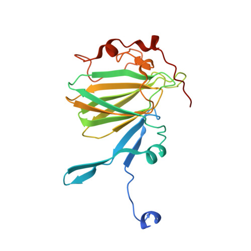
7PVI, 7PWB, 7PWH, 7PWI - PubMed Abstract:
The sugars streptose and dihydrohydroxystreptose (DHHS) are unique to the bacteria Streptomyces griseus and Coxiella burnetii, respectively. Streptose forms the central moiety of the antibiotic streptomycin, while DHHS is found in the O-antigen of the zoonotic pathogen C. burnetii. Biosynthesis of these sugars has been proposed to follow a similar path to that of TDP-rhamnose, catalyzed by the enzymes RmlA, RmlB, RmlC, and RmlD, but the exact mechanism is unclear. Streptose and DHHS biosynthesis unusually requires a ring contraction step that could be performed by orthologs of RmlC or RmlD. Genome sequencing of S. griseus and C. burnetii has identified StrM and CBU1838 proteins as RmlC orthologs in these respective species. Here, we demonstrate that both enzymes can perform the RmlC 3'',5'' double epimerization activity necessary to support TDP-rhamnose biosynthesis in vivo. This is consistent with the ring contraction step being performed on a double epimerized substrate. We further demonstrate that proton exchange is faster at the 3''-position than the 5''-position, in contrast to a previously studied ortholog. We additionally solved the crystal structures of CBU1838 and StrM in complex with TDP and show that they form an active site highly similar to those of the previously characterized enzymes RmlC, EvaD, and ChmJ. These results support the hypothesis that streptose and DHHS are biosynthesized using the TDP pathway and that an RmlD paralog most likely performs ring contraction following double epimerization. This work will support the elucidation of the full pathways for biosynthesis of these unique sugars.
- Living Systems Institute, University of Exeter, Exeter, United Kingdom; Department of Biosciences, University of Exeter, Exeter, United Kingdom.
Organizational Affiliation: