7PV6 | pdb_00007pv6

CARM1 in complex with EML734


Experimental Data Snapshot

  • Method: X-RAY DIFFRACTION
  • Resolution: 2.40 Å
  • R-Value Free: 
    0.237 (Depositor), 0.238 (DCC) 
  • R-Value Work: 
    0.191 (Depositor), 0.192 (DCC) 
  • R-Value Observed: 
    0.193 (Depositor) 

Starting Model: experimental
View more details

wwPDB Validation 3D Report Full Report

Validation slider image for 7PV6

Ligand Structure Quality Assessment 


This is version 1.3 of the entry. See complete history

Literature

Turning Nonselective Inhibitors of Type I Protein Arginine Methyltransferases into Potent and Selective Inhibitors of Protein Arginine Methyltransferase 4 through a Deconstruction-Reconstruction and Fragment-Growing Approach.

Iannelli, G.Milite, C.Marechal, N.Cura, V.Bonnefond, L.Troffer-Charlier, N.Feoli, A.Rescigno, D.Wang, Y.Cipriano, A.Viviano, M.Bedford, M.T.Cavarelli, J.Castellano, S.Sbardella, G.

(2022) J Med Chem 65: 11574-11606

  • DOI: https://doi.org/10.1021/acs.jmedchem.2c00252
  • Primary Citation Related Structures: 
    7NUD, 7NUE, 7P2R, 7PPQ, 7PPY, 7PU8, 7PUC, 7PUQ, 7PV6

  • PubMed Abstract: 

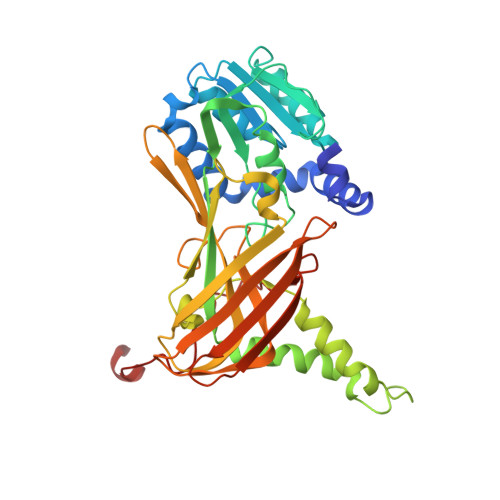
    Protein arginine methyltransferases (PRMTs) are important therapeutic targets, playing a crucial role in the regulation of many cellular processes and being linked to many diseases. Yet, there is still much to be understood regarding their functions and the biological pathways in which they are involved, as well as on the structural requirements that could drive the development of selective modulators of PRMT activity. Here we report a deconstruction-reconstruction approach that, starting from a series of type I PRMT inhibitors previously identified by us, allowed for the identification of potent and selective inhibitors of PRMT4, which regardless of the low cell permeability show an evident reduction of arginine methylation levels in MCF7 cells and a marked reduction of proliferation. We also report crystal structures with various PRMTs supporting the observed specificity and selectivity.


  • Organizational Affiliation
    • Department of Integrated Structural Biology, Institut de Génétique et de Biologie Moléculaire et Cellulaire, 67400 Illkirch, France.

Macromolecule Content 

  • Total Structure Weight: 170.01 kDa 
  • Atom Count: 11,684 
  • Modeled Residue Count: 1,410 
  • Deposited Residue Count: 1,472 
  • Unique protein chains: 1

Macromolecules

Find similar proteins by:|  3D Structure
Entity ID: 1
MoleculeChains  Sequence LengthOrganismDetailsImage
Histone-arginine methyltransferase CARM1
A, B, C, D
368Mus musculusMutation(s): 0 
Gene Names: Carm1Prmt4
EC: 2.1.1.319
UniProt & NIH Common Fund Data Resources
Find proteins for Q9WVG6 (Mus musculus)
Explore Q9WVG6 
Go to UniProtKB:  Q9WVG6
IMPC:  MGI:1913208
Entity Groups
Sequence Clusters30% Identity50% Identity70% Identity90% Identity95% Identity100% Identity
UniProt GroupQ9WVG6
Sequence Annotations
Expand
Reference Sequence

Small Molecules

Ligands 3 Unique
IDChains Name / Formula / InChI Key2D Diagram3D Interactions
LRZ
(Subject of Investigation/LOI)

Query on LRZ


Download:Ideal Coordinates CCD File
E [auth A],
L [auth B],
P [auth C],
U [auth D]
methyl 6-[3-[[~{N}-[[(2~{R},3~{S},4~{R},5~{R})-5-(6-aminopurin-9-yl)-3,4-bis(oxidanyl)oxolan-2-yl]methyl]carbamimidoyl]amino]propylcarbamoylamino]-4-oxidanyl-naphthalene-2-carboxylate
C27 H32 N10 O7
GJPQENHVEQGHGP-UMCMBGNQSA-N
PG4

Query on PG4


Download:Ideal Coordinates CCD File
T [auth C]TETRAETHYLENE GLYCOL
C8 H18 O5
UWHCKJMYHZGTIT-UHFFFAOYSA-N
EDO

Query on EDO


Download:Ideal Coordinates CCD File
F [auth A]
G [auth A]
H [auth A]
I [auth A]
J [auth A]
F [auth A],
G [auth A],
H [auth A],
I [auth A],
J [auth A],
K [auth A],
M [auth B],
N [auth B],
O [auth B],
Q [auth C],
R [auth C],
S [auth C]
1,2-ETHANEDIOL
C2 H6 O2
LYCAIKOWRPUZTN-UHFFFAOYSA-N
Binding Affinity Annotations 
IDSourceBinding Affinity
LRZ BindingDB:  7PV6 Kd: 663 (nM) from 1 assay(s)
IC50: 1.38e+4 (nM) from 1 assay(s)

Experimental Data & Validation

Experimental Data

  • Method: X-RAY DIFFRACTION
  • Resolution: 2.40 Å
  • R-Value Free:  0.237 (Depositor), 0.238 (DCC) 
  • R-Value Work:  0.191 (Depositor), 0.192 (DCC) 
  • R-Value Observed: 0.193 (Depositor) 
Space Group: P 21 21 2
Unit Cell:
Length ( Å )Angle ( ˚ )
a = 75α = 90
b = 99.519β = 90
c = 208.46γ = 90
Software Package:
Software NamePurpose
PHENIXrefinement
PDB_EXTRACTdata extraction
XDSdata reduction
XDSdata scaling
PHENIXphasing

Structure Validation

View Full Validation Report


Ligand Structure Quality Assessment 


Entry History 

& Funding Information

Deposition Data


Funding OrganizationLocationGrant Number
Not funded--

Revision History  (Full details and data files)

  • Version 1.0: 2022-04-06
    Type: Initial release
  • Version 1.1: 2022-05-11
    Changes: Database references
  • Version 1.2: 2022-09-21
    Changes: Database references
  • Version 1.3: 2024-01-31
    Changes: Data collection, Refinement description