7PU6

STRUCTURE OF ESTER-HYDROLASE EH7 FROM THE METAGENOME OF MARINE SEDIMENTS AT MILAZZO HARBOR (SICILY, ITALY) COMPLEXED WITH A DERIVATIVE OF OCTYL 4-NITROPHENYL HEXYLPHOSPHONATE


Experimental Data Snapshot

  • Method: X-RAY DIFFRACTION
  • Resolution: 2.92 Å
  • R-Value Free: 0.221 
  • R-Value Work: 0.183 
  • R-Value Observed: 0.185 

wwPDB Validation   3D Report Full Report


Ligand Structure Quality Assessment 


This is version 1.2 of the entry. See complete history


Literature

Crystal structure of a family VIII beta-lactamase fold hydrolase reveals the molecular mechanism for its broad substrate scope.

Cea-Rama, I.Coscolin, C.Gonzalez-Alfonso, J.L.Raj, J.Vasiljevic, M.Plou, F.J.Ferrer, M.Sanz-Aparicio, J.

(2022) FEBS J 289: 6714-6730

  • DOI: https://doi.org/10.1111/febs.16554
  • Primary Citation of Related Structures:  
    7PP3, 7PP8, 7PU6

  • PubMed Abstract: 

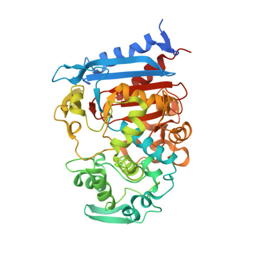
    Family VIII esterases present similarities to class C β-lactamases, which show nucleophilic serines located at the S-X-X-K motif instead of the G-X-S-X-G or G-D-S-(L) motif shown by other carboxylesterase families. Here, we report the crystal structure of a novel family VIII (subfamily VIII. I) esterase (EH 7 ; denaturing temperature, 52.6 ± 0.3 °C; pH optimum 7.0-9.0) to deepen its broad substrate range. Indeed, the analysis of the substrate specificity revealed its capacity to hydrolyse nitrocefin as a model chromogenic cephalosporin substrate (40.4 ± 11.4 units·g -1 ), and a large battery of 66 structurally different esters (up to 1730 min -1 ), including bis(2-hydroxyethyl)-terephthalate (241.7 ± 8.5 units·g -1 ) and the mycotoxin T-2 (1220 ± 52 units·g -1 ). It also showed acyltransferase activity through the synthesis of benzyl 3-oxobutanoate (40.4 ± 11.4 units·g -1 ) from benzyl alcohol and vinyl acetoacetate. Such a broad substrate scope is rare among family VIII esterases and lipolytic enzymes. Structural analyses of free and substrate-bound forms of this homooctamer esterase suggest that EH 7 presents a more opened and exposed S1 site having no steric hindrance for the entrance of substrates to the active site, more flexible R1, R2 and R3 regions allowing for the binding of a wide spectrum of substrates into the active site, and small residues in the conserved motif Y-X-X containing the catalytic Tyr enabling the entrance of large substrates. These unique structural elements in combination with docking experiments allowed us to gain valuable insights into the substrate specificity of this esterase and possible others belonging to family VIII.


  • Organizational Affiliation

    IQFR, CSIC, Madrid, Spain.


Macromolecules
Find similar proteins by:  (by identity cutoff)  |  3D Structure
Entity ID: 1
MoleculeChains Sequence LengthOrganismDetailsImage
Esterase
A, B, C, D, E
A, B, C, D, E, F, G, H
429metagenomeMutation(s): 0 
UniProt
Find proteins for A0A2K8JQ66 (uncultured bacterium)
Explore A0A2K8JQ66 
Go to UniProtKB:  A0A2K8JQ66
Entity Groups  
Sequence Clusters30% Identity50% Identity70% Identity90% Identity95% Identity100% Identity
UniProt GroupA0A2K8JQ66
Sequence Annotations
Expand
  • Reference Sequence
Small Molecules
Ligands 2 Unique
IDChains Name / Formula / InChI Key2D Diagram3D Interactions
P8K (Subject of Investigation/LOI)
Query on P8K

Download Ideal Coordinates CCD File 
I [auth A]
K [auth B]
L [auth C]
M [auth D]
N [auth E]
I [auth A],
K [auth B],
L [auth C],
M [auth D],
N [auth E],
O [auth F],
P [auth G],
Q [auth H]
hexyl(octoxy)phosphinic acid
C14 H31 O3 P
ITXAFDXPCLYZIG-UHFFFAOYSA-N
GOL
Query on GOL

Download Ideal Coordinates CCD File 
J [auth B]GLYCEROL
C3 H8 O3
PEDCQBHIVMGVHV-UHFFFAOYSA-N
Experimental Data & Validation

Experimental Data

  • Method: X-RAY DIFFRACTION
  • Resolution: 2.92 Å
  • R-Value Free: 0.221 
  • R-Value Work: 0.183 
  • R-Value Observed: 0.185 
  • Space Group: P 43 21 2
Unit Cell:
Length ( Å )Angle ( ˚ )
a = 149.541α = 90
b = 149.541β = 90
c = 321.718γ = 90
Software Package:
Software NamePurpose
REFMACrefinement
PDB_EXTRACTdata extraction
XDSdata reduction
XDSdata scaling
MOLREPphasing

Structure Validation

View Full Validation Report


Ligand Structure Quality Assessment 


Entry History & Funding Information

Deposition Data


Funding OrganizationLocationGrant Number
Spanish Ministry of Economy and CompetitivenessSpainProject BIO2016-76601-C3-3-R

Revision History  (Full details and data files)

  • Version 1.0: 2022-07-06
    Type: Initial release
  • Version 1.1: 2022-11-09
    Changes: Database references
  • Version 1.2: 2024-01-31
    Changes: Data collection, Refinement description