Structure, folding and flexibility of co-transcriptional RNA origami.
McRae, E.K.S., Rasmussen, H.O., Liu, J., Boggild, A., Nguyen, M.T.A., Sampedro Vallina, N., Boesen, T., Pedersen, J.S., Ren, G., Geary, C., Andersen, E.S.(2023) Nat Nanotechnol 18: 808-817
- PubMed: 36849548 Search on PubMedSearch on PubMed Central
- DOI: https://doi.org/10.1038/s41565-023-01321-6
- Primary Citation Related Structures:
7PTK, 7PTL, 7PTQ, 7PTS, 7QDU - PubMed Abstract:
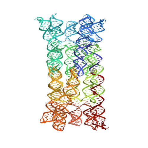
RNA origami is a method for designing RNA nanostructures that can self-assemble through co-transcriptional folding with applications in nanomedicine and synthetic biology. However, to advance the method further, an improved understanding of RNA structural properties and folding principles is required. Here we use cryogenic electron microscopy to study RNA origami sheets and bundles at sub-nanometre resolution revealing structural parameters of kissing-loop and crossover motifs, which are used to improve designs. In RNA bundle designs, we discover a kinetic folding trap that forms during folding and is only released after 10 h. Exploration of the conformational landscape of several RNA designs reveal the flexibility of helices and structural motifs. Finally, sheets and bundles are combined to construct a multidomain satellite shape, which is characterized by individual-particle cryo-electron tomography to reveal the domain flexibility. Together, the study provides a structural basis for future improvements to the design cycle of genetically encoded RNA nanodevices.
- Interdisciplinary Nanoscience Center (iNANO), Aarhus University, Aarhus, Denmark.
Organizational Affiliation: