7PRI

Carbonic Anhydrase from Schistosoma Mansoni in complex with clorsulon


Experimental Data Snapshot

  • Method: X-RAY DIFFRACTION
  • Resolution: 1.68 Å
  • R-Value Free: 0.195 
  • R-Value Work: 0.167 

wwPDB Validation   3D Report Full Report


Ligand Structure Quality Assessment 


This is version 1.2 of the entry. See complete history


Literature

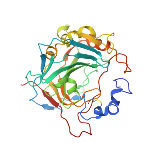
Inhibition of Schistosoma mansoni carbonic anhydrase by the antiparasitic drug clorsulon: X-ray crystallographic and in vitro studies.

Ferraroni, M.Angeli, A.Carradori, S.Supuran, C.T.

(2022) Acta Crystallogr D Struct Biol 78: 321-327

  • DOI: https://doi.org/10.1107/S2059798322000079
  • Primary Citation of Related Structures:  
    7PLF, 7PRI

  • PubMed Abstract: 

    Clorsulon is an anthelmintic drug that is clinically used against Fasciola hepatica. Due to the presence of two sulfonamide moieties in its core nucleus, which are well recognized as zinc-binding groups, it was proposed that it may be efficacious in the inhibition of parasite carbonic anhydrases (CAs). Proteomic analyses revealed the presence of CA in the tegument of Schistosoma mansoni, and recently the druggability of this target was explored by testing the inhibitory activities of several sulfonamide-based derivatives. According to the principles of drug repurposing, the aim was to demonstrate a putative new mechanism of action of clorsulon and thus widen its antiparasitic spectrum. For this purpose, the inhibitory activity and isoform selectivity of clorsulon was studied using human CA I and S. mansoni CA, revealing different modes of binding of clorsulon that explain its inhibitory potency against the two enzymes. The information obtained in this study could be crucial in the design of more active and selective derivatives.


  • Organizational Affiliation

    Dipartimento di Chimica `Ugo Schiff', University of Florence, Via della Lastruccia 3, 50019 Sesto Fiorentino, Florence, Italy.


Macromolecules
Find similar proteins by:  (by identity cutoff)  |  3D Structure
Entity ID: 1
MoleculeChains Sequence LengthOrganismDetailsImage
Carbonic anhydraseA [auth AAA],
B [auth BBB]
280Schistosoma mansoniMutation(s): 0 
EC: 4.2.1.1
UniProt
Find proteins for A0A3Q0KSG2 (Schistosoma mansoni)
Explore A0A3Q0KSG2 
Go to UniProtKB:  A0A3Q0KSG2
Entity Groups  
Sequence Clusters30% Identity50% Identity70% Identity90% Identity95% Identity100% Identity
UniProt GroupA0A3Q0KSG2
Sequence Annotations
Expand
  • Reference Sequence
Small Molecules
Ligands 4 Unique
IDChains Name / Formula / InChI Key2D Diagram3D Interactions
7TI (Subject of Investigation/LOI)
Query on 7TI

Download Ideal Coordinates CCD File 
F [auth AAA],
L [auth BBB]
4-azanyl-6-[1,2,2-tris(chloranyl)ethenyl]benzene-1,3-disulfonamide
C8 H8 Cl3 N3 O4 S2
QOVTVIYTBRHADL-UHFFFAOYSA-N
NAG (Subject of Investigation/LOI)
Query on NAG

Download Ideal Coordinates CCD File 
D [auth AAA],
E [auth AAA],
I [auth BBB],
J [auth BBB],
K [auth BBB]
2-acetamido-2-deoxy-beta-D-glucopyranose
C8 H15 N O6
OVRNDRQMDRJTHS-FMDGEEDCSA-N
GOL
Query on GOL

Download Ideal Coordinates CCD File 
G [auth BBB]GLYCEROL
C3 H8 O3
PEDCQBHIVMGVHV-UHFFFAOYSA-N
ZN
Query on ZN

Download Ideal Coordinates CCD File 
C [auth AAA],
H [auth BBB]
ZINC ION
Zn
PTFCDOFLOPIGGS-UHFFFAOYSA-N
Experimental Data & Validation

Experimental Data

  • Method: X-RAY DIFFRACTION
  • Resolution: 1.68 Å
  • R-Value Free: 0.195 
  • R-Value Work: 0.167 
  • Space Group: P 32 2 1
Unit Cell:
Length ( Å )Angle ( ˚ )
a = 103.8α = 90
b = 103.8β = 90
c = 132.9γ = 120
Software Package:
Software NamePurpose
REFMACrefinement
XDSdata reduction
XSCALEdata scaling
REFMACphasing

Structure Validation

View Full Validation Report


Ligand Structure Quality Assessment 


Entry History & Funding Information

Deposition Data


Funding OrganizationLocationGrant Number
Not funded--

Revision History  (Full details and data files)

  • Version 1.0: 2022-10-05
    Type: Initial release
  • Version 1.1: 2023-04-19
    Changes: Database references
  • Version 1.2: 2024-02-07
    Changes: Data collection, Refinement description