7PLB

Caulobacter crescentus xylonolactonase with D-xylose


Experimental Data Snapshot

  • Method: X-RAY DIFFRACTION
  • Resolution: 1.73 Å
  • R-Value Free: 0.218 
  • R-Value Work: 0.181 
  • R-Value Observed: 0.182 

wwPDB Validation   3D Report Full Report


Ligand Structure Quality Assessment 


This is version 1.2 of the entry. See complete history


Literature

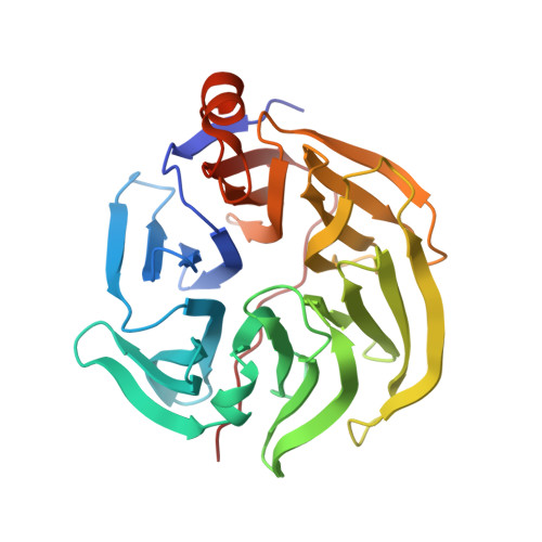
Three-dimensional structure of xylonolactonase from Caulobacter crescentus: A mononuclear iron enzyme of the 6-bladed beta-propeller hydrolase family.

Paakkonen, J.Hakulinen, N.Andberg, M.Koivula, A.Rouvinen, J.

(2022) Protein Sci 31: 371-383

  • DOI: https://doi.org/10.1002/pro.4229
  • Primary Citation of Related Structures:  
    7PLB, 7PLC, 7PLD

  • PubMed Abstract: 

    Xylonolactonase Cc XylC from Caulobacter crescentus catalyzes the hydrolysis of the intramolecular ester bond of d-xylonolactone. We have determined crystal structures of Cc XylC in complex with d-xylonolactone isomer analogues d-xylopyranose and (r)-(+)-4-hydroxy-2-pyrrolidinone at high resolution. Cc XylC has a 6-bladed β-propeller architecture, which contains a central open channel having the active site at one end. According to our previous native mass spectrometry studies, Cc XylC is able to specifically bind Fe 2+ . The crystal structures, presented here, revealed an active site bound metal ion with an octahedral binding geometry. The side chains of three amino acid residues, Glu18, Asn146, and Asp196, which participate in binding of metal ion are located in the same plane. The solved complex structures allowed suggesting a reaction mechanism for intramolecular ester bond hydrolysis in which the major contribution for catalysis arises from the carbonyl oxygen coordination of the xylonolactone substrate to the Fe 2+ . The structure of Cc XylC was compared with eight other ester hydrolases of the β-propeller hydrolase family. The previously published crystal structures of other β-propeller hydrolases contain either Ca 2+ , Mg 2+ , or Zn 2+ and show clear similarities in ligand and metal ion binding geometries to that of Cc XylC. It would be interesting to reinvestigate the metal binding specificity of these enzymes and clarify whether they are also able to use Fe 2+ as a catalytic metal. This could further expand our understanding of utilization of Fe 2+ not only in oxidative enzymes but also in hydrolases.


  • Organizational Affiliation

    Department of Chemistry, University of Eastern Finland, Joensuu, Finland.


Macromolecules
Find similar proteins by:  (by identity cutoff)  |  3D Structure
Entity ID: 1
MoleculeChains Sequence LengthOrganismDetailsImage
Smp-30/Cgr1 family protein
A, B
290Caulobacter vibrioides CB15Mutation(s): 0 
Gene Names: CC_0820
EC: 3.1.1.68
UniProt
Find proteins for Q9A9Z1 (Caulobacter vibrioides (strain ATCC 19089 / CB15))
Explore Q9A9Z1 
Go to UniProtKB:  Q9A9Z1
Entity Groups  
Sequence Clusters30% Identity50% Identity70% Identity90% Identity95% Identity100% Identity
UniProt GroupQ9A9Z1
Sequence Annotations
Expand
  • Reference Sequence
Small Molecules
Ligands 4 Unique
IDChains Name / Formula / InChI Key2D Diagram3D Interactions
XYS (Subject of Investigation/LOI)
Query on XYS

Download Ideal Coordinates CCD File 
J [auth B]alpha-D-xylopyranose
C5 H10 O5
SRBFZHDQGSBBOR-LECHCGJUSA-N
XYP (Subject of Investigation/LOI)
Query on XYP

Download Ideal Coordinates CCD File 
E [auth A]
F [auth A]
G [auth A]
I [auth B]
K [auth B]
E [auth A],
F [auth A],
G [auth A],
I [auth B],
K [auth B],
L [auth B]
beta-D-xylopyranose
C5 H10 O5
SRBFZHDQGSBBOR-KKQCNMDGSA-N
SO4
Query on SO4

Download Ideal Coordinates CCD File 
D [auth A]SULFATE ION
O4 S
QAOWNCQODCNURD-UHFFFAOYSA-L
FE2 (Subject of Investigation/LOI)
Query on FE2

Download Ideal Coordinates CCD File 
C [auth A],
H [auth B]
FE (II) ION
Fe
CWYNVVGOOAEACU-UHFFFAOYSA-N
Experimental Data & Validation

Experimental Data

  • Method: X-RAY DIFFRACTION
  • Resolution: 1.73 Å
  • R-Value Free: 0.218 
  • R-Value Work: 0.181 
  • R-Value Observed: 0.182 
  • Space Group: C 2 2 21
Unit Cell:
Length ( Å )Angle ( ˚ )
a = 85.84α = 90
b = 170.95β = 90
c = 79.14γ = 90
Software Package:
Software NamePurpose
PHENIXrefinement
GDAdata collection
xia2data reduction
DIALSdata scaling
Cootmodel building
MR-Rosettaphasing
PHASERphasing

Structure Validation

View Full Validation Report


Ligand Structure Quality Assessment 


Entry History & Funding Information

Deposition Data


Funding OrganizationLocationGrant Number
Academy of FinlandFinland118573
Academy of FinlandFinland287241
Academy of FinlandFinland288677
Academy of FinlandFinland322610

Revision History  (Full details and data files)

  • Version 1.0: 2021-11-24
    Type: Initial release
  • Version 1.1: 2022-02-16
    Changes: Data collection, Database references
  • Version 1.2: 2024-01-31
    Changes: Data collection, Refinement description