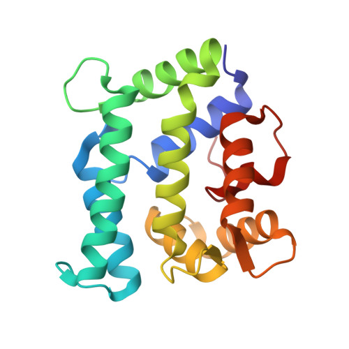
The X-ray structure of juvenile hormone diol kinase from the silkworm Bombyx mori.
Guo, J., Keegan, R.M., Rigden, D.J., Erskine, P.T., Wood, S.P., Li, S., Cooper, J.B.(2021) Acta Crystallogr F Struct Biol Commun 77: 465-472
- PubMed: 34866602 Search on PubMedSearch on PubMed Central
- DOI: https://doi.org/10.1107/S2053230X21012012
- Primary Citation Related Structures:
7PJD - PubMed Abstract:
Insect juvenile hormones (JHs) are a family of sesquiterpenoid molecules that are secreted into the haemolymph. JHs have multiple roles in insect development, metamorphosis and sexual maturation. A number of pesticides work by chemically mimicking JHs, thus preventing insects from developing and reproducing normally. The haemolymph levels of JH are governed by the rates of its biosynthesis and degradation. One enzyme involved in JH catabolism is JH diol kinase (JHDK), which uses ATP (or GTP) to phosphorylate JH diol to JH diol phosphate, which can be excreted. The X-ray structure of JHDK from the silkworm Bombyx mori has been determined at a resolution of 2.0 Å with an R factor of 19.0% and an R free of 24.8%. The structure possesses three EF-hand motifs which are occupied by calcium ions. This is in contrast to the recently reported structure of the JHDK-like-2 protein from B. mori (PDB entry 6kth), which possessed only one calcium ion. Since JHDK is known to be inhibited by calcium ions, it is likely that our structure represents the calcium-inhibited form of the enzyme. The electrostatic surface of the protein suggests a binding site for the triphosphate of ATP close to the N-terminal end of the molecule in a cavity between the N- and C-terminal domains. Superposition with a number of calcium-activated photoproteins suggests that there may be parallels between the binding of JH diol to JHDK and the binding of luciferin to aequorin.
- Division of Medicine, UCL, Gower Street, London WC1E 6BT, United Kingdom.
Organizational Affiliation: