


Crystal structure of Bacillus subtilis PabB, component 1.
Rooms, L.D., Race, P.R., Devine, A., Willis, C.L., Back, C.R., Burton, N., Sudol, A.To be published.
Experimental Data Snapshot
Starting Model: other
View more details
Entity ID: 1 | |||||
|---|---|---|---|---|---|
| Molecule | Chains | Sequence Length | Organism | Details | Image |
| Aminodeoxychorismate synthase component 1 | A [auth AAA], B [auth BBB], C [auth CCC], D [auth DDD] | 489 | Bacillus subtilis subsp. subtilis str. 168 | Mutation(s): 0 Gene Names: pabB, pab, BSU00740 EC: 2.6.1.85 | 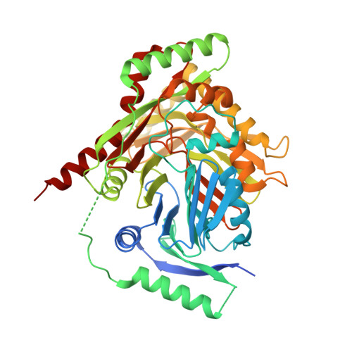 |
UniProt | |||||
Entity Groups | |||||
| Sequence Clusters | 30% Identity50% Identity70% Identity90% Identity95% Identity100% Identity | ||||
| UniProt Group | P28820 | ||||
Sequence AnnotationsExpand | |||||
Reference Sequence | |||||
| Ligands 2 Unique | |||||
|---|---|---|---|---|---|
| ID | Chains | Name / Formula / InChI Key | 2D Diagram | 3D Interactions | |
| TRP (Subject of Investigation/LOI) Download:Ideal Coordinates CCD File | F [auth AAA], G [auth BBB], I [auth CCC], K [auth DDD] | TRYPTOPHAN C11 H12 N2 O2 QIVBCDIJIAJPQS-VIFPVBQESA-N |  | ||
| MG (Subject of Investigation/LOI) Download:Ideal Coordinates CCD File | E [auth AAA], H [auth BBB], J [auth CCC], L [auth DDD] | MAGNESIUM ION Mg JLVVSXFLKOJNIY-UHFFFAOYSA-N |  | ||
| Length ( Å ) | Angle ( ˚ ) |
|---|---|
| a = 51.084 | α = 90 |
| b = 170.698 | β = 90 |
| c = 224.99 | γ = 90 |
| Software Name | Purpose |
|---|---|
| REFMAC | refinement |
| xia2 | data reduction |
| DIALS | data scaling |
| PHASER | phasing |
| Funding Organization | Location | Grant Number |
|---|---|---|
| Other government | United Kingdom | -- |


