7PHH

Human voltage-gated potassium channel Kv3.1 (apo condition)


Experimental Data Snapshot

  • Method: ELECTRON MICROSCOPY
  • Resolution: 3.20 Å
  • Aggregation State: PARTICLE 
  • Reconstruction Method: SINGLE PARTICLE 

wwPDB Validation   3D Report Full Report


This is version 1.1 of the entry. See complete history


Literature

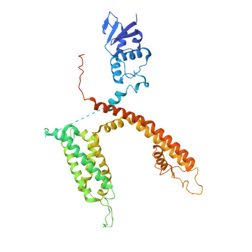
Cryo-EM structure of the human Kv3.1 channel reveals gating control by the cytoplasmic T1 domain.

Chi, G.Liang, Q.Sridhar, A.Cowgill, J.B.Sader, K.Radjainia, M.Qian, P.Castro-Hartmann, P.Venkaya, S.Singh, N.K.McKinley, G.Fernandez-Cid, A.Mukhopadhyay, S.M.M.Burgess-Brown, N.A.Delemotte, L.Covarrubias, M.Durr, K.L.

(2022) Nat Commun 13: 4087-4087

  • DOI: https://doi.org/10.1038/s41467-022-29594-w
  • Primary Citation of Related Structures:  
    7PHH, 7PHI, 7PHK, 7PHL

  • PubMed Abstract: 

    Kv3 channels have distinctive gating kinetics tailored for rapid repolarization in fast-spiking neurons. Malfunction of this process due to genetic variants in the KCNC1 gene causes severe epileptic disorders, yet the structural determinants for the unusual gating properties remain elusive. Here, we present cryo-electron microscopy structures of the human Kv3.1a channel, revealing a unique arrangement of the cytoplasmic tetramerization domain T1 which facilitates interactions with C-terminal axonal targeting motif and key components of the gating machinery. Additional interactions between S1/S2 linker and turret domain strengthen the interface between voltage sensor and pore domain. Supported by molecular dynamics simulations, electrophysiological and mutational analyses, we identify several residues in the S4/S5 linker which influence the gating kinetics and an electrostatic interaction between acidic residues in α6 of T1 and R449 in the pore-flanking S6T helices. These findings provide insights into gating control and disease mechanisms and may guide strategies for the design of pharmaceutical drugs targeting Kv3 channels.


  • Organizational Affiliation

    Centre for Medicines Discovery, Nuffield Department of Medicine, University of Oxford, Roosevelt Drive, Oxford, OX3 7DQ, UK.


Macromolecules
Find similar proteins by:  (by identity cutoff)  |  3D Structure
Entity ID: 1
MoleculeChains Sequence LengthOrganismDetailsImage
Potassium voltage-gated channel, Shaw-related subfamily, member 1A,
B [auth C],
C [auth B],
D
518Homo sapiensMutation(s): 0 
Gene Names: KCNC1
Membrane Entity: Yes 
UniProt & NIH Common Fund Data Resources
Find proteins for P48547 (Homo sapiens)
Explore P48547 
Go to UniProtKB:  P48547
PHAROS:  P48547
GTEx:  ENSG00000129159 
Entity Groups  
Sequence Clusters30% Identity50% Identity70% Identity90% Identity95% Identity100% Identity
UniProt GroupP48547
Sequence Annotations
Expand
  • Reference Sequence
Small Molecules
Ligands 3 Unique
IDChains Name / Formula / InChI Key2D Diagram3D Interactions
PCF
Query on PCF

Download Ideal Coordinates CCD File 
E [auth A]
F [auth A]
L [auth C]
M [auth C]
P [auth B]
E [auth A],
F [auth A],
L [auth C],
M [auth C],
P [auth B],
Q [auth B],
S [auth D],
T [auth D]
1,2-DIACYL-SN-GLYCERO-3-PHOSHOCHOLINE
C40 H80 N O8 P
KILNVBDSWZSGLL-KXQOOQHDSA-N
ZN
Query on ZN

Download Ideal Coordinates CCD File 
G [auth A],
N [auth C],
O [auth B],
R [auth D]
ZINC ION
Zn
PTFCDOFLOPIGGS-UHFFFAOYSA-N
K
Query on K

Download Ideal Coordinates CCD File 
H [auth A],
I [auth A],
J [auth A],
K [auth A]
POTASSIUM ION
K
NPYPAHLBTDXSSS-UHFFFAOYSA-N
Experimental Data & Validation

Experimental Data

  • Method: ELECTRON MICROSCOPY
  • Resolution: 3.20 Å
  • Aggregation State: PARTICLE 
  • Reconstruction Method: SINGLE PARTICLE 

Structure Validation

View Full Validation Report


Entry History & Funding Information

Deposition Data


Funding OrganizationLocationGrant Number
Wellcome TrustUnited Kingdom106169/Z/14/Z

Revision History  (Full details and data files)

  • Version 1.0: 2022-03-02
    Type: Initial release
  • Version 1.1: 2022-08-10
    Changes: Database references