


Crystal structure of Phenazine 1-carboxylic acid decarboxylase from Mycobacterium fortuitum
Gahloth, D., Leys, D.To be published.
Experimental Data Snapshot
Starting Model: experimental
View more details
Entity ID: 1 | |||||
|---|---|---|---|---|---|
| Molecule | Chains | Sequence Length | Organism | Details | Image |
| UbiD family decarboxylase | 479 | Mycolicibacterium fortuitum | Mutation(s): 0 Gene Names: XA26_16650 | 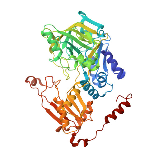 | |
UniProt | |||||
Find proteins for A0A0N9Y7U2 (Mycolicibacterium fortuitum) Explore A0A0N9Y7U2 Go to UniProtKB: A0A0N9Y7U2 | |||||
Entity Groups | |||||
| Sequence Clusters | 30% Identity50% Identity70% Identity90% Identity95% Identity100% Identity | ||||
| UniProt Group | A0A0N9Y7U2 | ||||
Sequence AnnotationsExpand | |||||
Reference Sequence | |||||
Entity ID: 2 | |||||
|---|---|---|---|---|---|
| Molecule | Chains | Sequence Length | Organism | Details | Image |
| UNK | 5 | unidentified | Mutation(s): 0 | 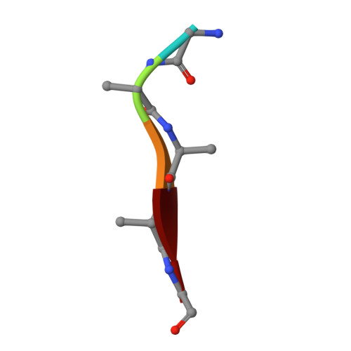 | |
| Ligands 3 Unique | |||||
|---|---|---|---|---|---|
| ID | Chains | Name / Formula / InChI Key | 2D Diagram | 3D Interactions | |
| 4LU (Subject of Investigation/LOI) Download:Ideal Coordinates CCD File | D [auth A] | 1-deoxy-5-O-phosphono-1-(3,3,4,5-tetramethyl-9,11-dioxo-2,3,8,9,10,11-hexahydro-7H-quinolino[1,8-fg]pteridin-12-ium-7-y
l)-D-ribitol C22 H30 N4 O9 P KOUJZPGFPGLHCZ-IYOUNJFTSA-O |  | ||
| MN Download:Ideal Coordinates CCD File | C [auth A] | MANGANESE (II) ION Mn WAEMQWOKJMHJLA-UHFFFAOYSA-N |  | ||
| NA Download:Ideal Coordinates CCD File | E [auth A] | SODIUM ION Na FKNQFGJONOIPTF-UHFFFAOYSA-N |  | ||
| Length ( Å ) | Angle ( ˚ ) |
|---|---|
| a = 124.696 | α = 90 |
| b = 124.696 | β = 90 |
| c = 68.846 | γ = 120 |
| Software Name | Purpose |
|---|---|
| REFMAC | refinement |
| PDB_EXTRACT | data extraction |
| xia2 | data reduction |
| XDS | data scaling |
| PHASER | phasing |
| Funding Organization | Location | Grant Number |
|---|---|---|
| European Research Council (ERC) | European Union | -- |


