7PAG | pdb_00007pag

The pore conformation of lymphocyte perforin


Experimental Data Snapshot

  • Method: ELECTRON MICROSCOPY
  • Resolution: 4.00 Å
  • Aggregation State: PARTICLE 
  • Reconstruction Method: SINGLE PARTICLE 

wwPDB Validation 3D Report Full Report

Validation slider image for 7PAG

This is version 1.2 of the entry. See complete history

Literature

The pore conformation of lymphocyte perforin.

Ivanova, M.E.Lukoyanova, N.Malhotra, S.Topf, M.Trapani, J.A.Voskoboinik, I.Saibil, H.R.

(2022) Sci Adv 8: eabk3147-eabk3147

  • DOI: https://doi.org/10.1126/sciadv.abk3147
  • Primary Citation Related Structures: 
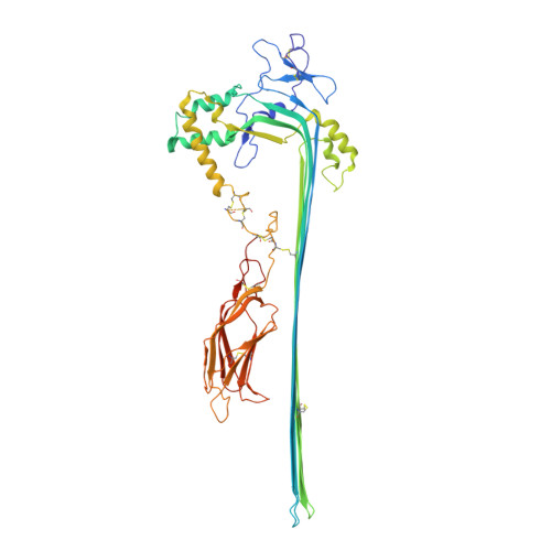
    7PAG

  • PubMed Abstract: 

    Perforin is a pore-forming protein that facilitates rapid killing of pathogen-infected or cancerous cells by the immune system. Perforin is released from cytotoxic lymphocytes, together with proapoptotic granzymes, to bind to a target cell membrane where it oligomerizes and forms pores. The pores allow granzyme entry, which rapidly triggers the apoptotic death of the target cell. Here, we present a 4-Å resolution cryo-electron microscopy structure of the perforin pore, revealing previously unidentified inter- and intramolecular interactions stabilizing the assembly. During pore formation, the helix-turn-helix motif moves away from the bend in the central β sheet to form an intermolecular contact. Cryo-electron tomography shows that prepores form on the membrane surface with minimal conformational changes. Our findings suggest the sequence of conformational changes underlying oligomerization and membrane insertion, and explain how several pathogenic mutations affect function.


  • Organizational Affiliation
    • Institute of Structural and Molecular Biology, Birkbeck, University of London, Malet St, London WC1E 7HX, UK.

Macromolecule Content 

  • Total Structure Weight: 61.17 kDa 
  • Atom Count: 4,040 
  • Modeled Residue Count: 509 
  • Deposited Residue Count: 540 
  • Unique protein chains: 1

Macromolecules

Find similar proteins by:|  3D Structure
Entity ID: 1
MoleculeChains  Sequence LengthOrganismDetailsImage
Perforin-1540Mus musculusMutation(s): 0 
Gene Names: Prf1Pfp
Membrane Entity: Yes 
UniProt & NIH Common Fund Data Resources
Find proteins for P10820 (Mus musculus)
Explore P10820 
Go to UniProtKB:  P10820
IMPC:  MGI:97551
Entity Groups
Sequence Clusters30% Identity50% Identity70% Identity90% Identity95% Identity100% Identity
UniProt GroupP10820
Glycosylation
Glycosylation Sites: 1Go to GlyGen: P10820-1
Sequence Annotations
Expand
Reference Sequence

Experimental Data & Validation

Experimental Data

  • Method: ELECTRON MICROSCOPY
  • Resolution: 4.00 Å
  • Aggregation State: PARTICLE 
  • Reconstruction Method: SINGLE PARTICLE 

Structure Validation

View Full Validation Report


Entry History 

& Funding Information

Deposition Data


Funding OrganizationLocationGrant Number
Wellcome TrustUnited Kingdom106249/Z/14/Z
Wellcome TrustUnited Kingdom209250/Z/17/Z
Wellcome TrustUnited Kingdom208398/Z/17/Z
European Research Council (ERC)United Kingdom294408
Wellcome TrustUnited Kingdom202679/Z/16/Z
Wellcome TrustUnited Kingdom206166/Z/17/Z

Revision History  (Full details and data files)

  • Version 1.0: 2022-02-16
    Type: Initial release
  • Version 1.1: 2022-02-23
    Changes: Database references
  • Version 1.2: 2024-11-13
    Changes: Data collection, Structure summary