Validation of a small molecule inhibitor of PDE6D-RAS interaction with favorable anti-leukemic effects.
Canovas Nunes, S., De Vita, S., Anighoro, A., Autelitano, F., Beaumont, E., Klingbeil, P., McGuinness, M., Duvert, B., Harris, C., Yang, L., Pokharel, S.P., Chen, C.W., Ermann, M., Williams, D.A., Xu, H.(2022) Blood Cancer J 12: 64-64
- PubMed: 35422065 Search on PubMedSearch on PubMed Central
- DOI: https://doi.org/10.1038/s41408-022-00663-z
- Primary Citation Related Structures:
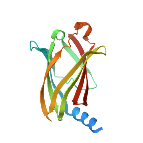
7PAC, 7PAD, 7PAE - PubMed Abstract:
RAS mutations prevalent in high-risk leukemia have been linked to relapse and chemotherapy resistance. Efforts to directly target RAS proteins have been largely unsuccessful. However, since RAS-mediated transformation is dependent on signaling through the RAS-related C3 botulinum toxin substrate (RAC) small GTPase, we hypothesized that targeting RAC may be an effective therapeutic approach in RAS mutated tumors. Here we describe multiple small molecules capable of inhibiting RAC activation in acute lymphoblastic leukemia cell lines. One of these, DW0254, also demonstrates promising anti-leukemic activity in RAS-mutated cells. Using chemical proteomics and biophysical methods, we identified the hydrophobic pocket of phosphodiester 6 subunit delta (PDE6D), a known RAS chaperone, as a target for this compound. Inhibition of RAS localization to the plasma membrane upon DW0254 treatment is associated with RAC inhibition through a phosphatidylinositol-3-kinase/AKT-dependent mechanism. Our findings provide new insights into the importance of PDE6D-mediated transport for RAS-dependent RAC activation and leukemic cell survival.
- Division of Hematology/Oncology, Boston Children's Hospital, Harvard Medical School, Boston, MA, USA.
Organizational Affiliation: