7P7P

Crystal structure of ERAP2 aminopeptidase in complex with phosphinic pseudotripeptide((1R)-1-Amino-3-phenylpropyl){(2S)-3-[((2S)-1-amino-1-oxo-3-phenylpropan-2-yl)amino]-2-{[3-(2-hydroxyphenyl)-isoxazol-5-yl]methyl}-3-oxopropyl}phosphinic acid


Experimental Data Snapshot

  • Method: X-RAY DIFFRACTION
  • Resolution: 3.00 Å
  • R-Value Free: 0.231 
  • R-Value Work: 0.180 
  • R-Value Observed: 0.182 

wwPDB Validation   3D Report Full Report


Ligand Structure Quality Assessment 


This is version 1.2 of the entry. See complete history


Literature

Inhibitor-Dependent Usage of the S1' Specificity Pocket of ER Aminopeptidase 2.

Mpakali, A.Georgiadis, D.Stratikos, E.Giastas, P.

(2022) ACS Med Chem Lett 13: 218-224

  • DOI: https://doi.org/10.1021/acsmedchemlett.1c00582
  • Primary Citation of Related Structures:  
    7P7P, 7PFS

  • PubMed Abstract: 

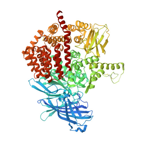
    Endoplasmic reticulum aminopeptidase 2 (ERAP2) is an intracellular enzyme involved in the processing of antigenic peptides intended for presentation by major histocompatibility complex class I (MHCI) molecules. Because of its role in regulating immune responses, ERAP2 is an emerging pharmacological target. Phosphinic pseudopeptides are potent transition-state analogue inhibitors of ERAP2. Previous structure-activity studies have revealed a complex but ambiguous relationship between the occupation of putative specificity pockets and the inhibitor efficacy. To address these problems, we solved crystal structures of ERAP2 in complex with two phosphinic pseudotripeptide inhibitors. Both compounds are found in the catalytic site in a canonical orientation for transition-state analogues and utilize the S1 and S2' pockets in a similar fashion. Strikingly, their P1' side chains exhibit different orientations and make interactions with distinct shallow pockets near the ERAP2 active site. These structures suggest that S1' pocket usage in ERAP2 may be inhibitor-dependent and constitute useful starting templates for the further optimization of this class of compounds.


  • Organizational Affiliation

    National Centre for Scientific Research Demokritos, Agia Paraskevi, Athens 15341, Greece.


Macromolecules
Find similar proteins by:  (by identity cutoff)  |  3D Structure
Entity ID: 1
MoleculeChains Sequence LengthOrganismDetailsImage
Endoplasmic reticulum aminopeptidase 2
A, B
962Homo sapiensMutation(s): 1 
Gene Names: ERAP2LRAP
EC: 3.4.11
UniProt & NIH Common Fund Data Resources
Find proteins for Q6P179 (Homo sapiens)
Explore Q6P179 
Go to UniProtKB:  Q6P179
PHAROS:  Q6P179
GTEx:  ENSG00000164308 
Entity Groups  
Sequence Clusters30% Identity50% Identity70% Identity90% Identity95% Identity100% Identity
UniProt GroupQ6P179
Sequence Annotations
Expand
  • Reference Sequence
Oligosaccharides

Help

Entity ID: 2
MoleculeChains Length2D Diagram Glycosylation3D Interactions
alpha-D-mannopyranose-(1-3)-beta-D-mannopyranose-(1-4)-2-acetamido-2-deoxy-beta-D-glucopyranose-(1-4)-2-acetamido-2-deoxy-beta-D-glucopyranose
C
4N-Glycosylation
Glycosylation Resources
GlyTouCan:  G81315DD
GlyCosmos:  G81315DD
GlyGen:  G81315DD
Entity ID: 3
MoleculeChains Length2D Diagram Glycosylation3D Interactions
alpha-D-mannopyranose-(1-6)-beta-D-mannopyranose-(1-4)-2-acetamido-2-deoxy-beta-D-glucopyranose-(1-4)-2-acetamido-2-deoxy-beta-D-glucopyranose
D
4N-Glycosylation
Glycosylation Resources
GlyTouCan:  G22573RC
GlyCosmos:  G22573RC
GlyGen:  G22573RC
Entity ID: 4
MoleculeChains Length2D Diagram Glycosylation3D Interactions
2-acetamido-2-deoxy-beta-D-glucopyranose-(1-4)-2-acetamido-2-deoxy-beta-D-glucopyranose
E, H, I
2N-Glycosylation
Glycosylation Resources
GlyTouCan:  G42666HT
GlyCosmos:  G42666HT
GlyGen:  G42666HT
Entity ID: 5
MoleculeChains Length2D Diagram Glycosylation3D Interactions
beta-D-mannopyranose-(1-4)-2-acetamido-2-deoxy-beta-D-glucopyranose-(1-4)-2-acetamido-2-deoxy-beta-D-glucopyranose
F
3N-Glycosylation
Glycosylation Resources
GlyTouCan:  G15407YE
GlyCosmos:  G15407YE
GlyGen:  G15407YE
Entity ID: 6
MoleculeChains Length2D Diagram Glycosylation3D Interactions
alpha-D-mannopyranose-(1-3)-[alpha-D-mannopyranose-(1-6)]beta-D-mannopyranose-(1-4)-2-acetamido-2-deoxy-beta-D-glucopyranose-(1-4)-2-acetamido-2-deoxy-beta-D-glucopyranose
G
5N-Glycosylation
Glycosylation Resources
GlyTouCan:  G22768VO
GlyCosmos:  G22768VO
GlyGen:  G22768VO
Small Molecules
Ligands 9 Unique
IDChains Name / Formula / InChI Key2D Diagram3D Interactions
62S
Query on 62S

Download Ideal Coordinates CCD File 
CA [auth B],
J [auth A]
[(2~{S})-3-[[(2~{S})-1-azanyl-1-oxidanylidene-3-phenyl-propan-2-yl]amino]-2-[[3-(2-hydroxyphenyl)-1,2-oxazol-5-yl]methyl]-3-oxidanylidene-propyl]-[(1~{R})-1-azanyl-3-phenyl-propyl]phosphinic acid
C31 H35 N4 O6 P
GSWUIYHGTPVQDP-FNHKZSGOSA-N
NAG
Query on NAG

Download Ideal Coordinates CCD File 
DA [auth B]
EA [auth B]
FA [auth B]
GA [auth B]
HA [auth B]
DA [auth B],
EA [auth B],
FA [auth B],
GA [auth B],
HA [auth B],
IA [auth B],
K [auth A],
L [auth A],
M [auth A],
N [auth A]
2-acetamido-2-deoxy-beta-D-glucopyranose
C8 H15 N O6
OVRNDRQMDRJTHS-FMDGEEDCSA-N
MES
Query on MES

Download Ideal Coordinates CCD File 
R [auth A]2-(N-MORPHOLINO)-ETHANESULFONIC ACID
C6 H13 N O4 S
SXGZJKUKBWWHRA-UHFFFAOYSA-N
PGE
Query on PGE

Download Ideal Coordinates CCD File 
Q [auth A]TRIETHYLENE GLYCOL
C6 H14 O4
ZIBGPFATKBEMQZ-UHFFFAOYSA-N
PEG
Query on PEG

Download Ideal Coordinates CCD File 
O [auth A],
P [auth A]
DI(HYDROXYETHYL)ETHER
C4 H10 O3
MTHSVFCYNBDYFN-UHFFFAOYSA-N
PDO
Query on PDO

Download Ideal Coordinates CCD File 
JA [auth B],
U [auth A]
1,3-PROPANDIOL
C3 H8 O2
YPFDHNVEDLHUCE-UHFFFAOYSA-N
IMD
Query on IMD

Download Ideal Coordinates CCD File 
W [auth A]IMIDAZOLE
C3 H5 N2
RAXXELZNTBOGNW-UHFFFAOYSA-O
ZN
Query on ZN

Download Ideal Coordinates CCD File 
BA [auth A],
KA [auth B]
ZINC ION
Zn
PTFCDOFLOPIGGS-UHFFFAOYSA-N
EDO
Query on EDO

Download Ideal Coordinates CCD File 
AA [auth A]
S [auth A]
T [auth A]
V [auth A]
X [auth A]
AA [auth A],
S [auth A],
T [auth A],
V [auth A],
X [auth A],
Y [auth A],
Z [auth A]
1,2-ETHANEDIOL
C2 H6 O2
LYCAIKOWRPUZTN-UHFFFAOYSA-N
Experimental Data & Validation

Experimental Data

  • Method: X-RAY DIFFRACTION
  • Resolution: 3.00 Å
  • R-Value Free: 0.231 
  • R-Value Work: 0.180 
  • R-Value Observed: 0.182 
  • Space Group: P 1 21 1
Unit Cell:
Length ( Å )Angle ( ˚ )
a = 75.342α = 90
b = 134.618β = 90.478
c = 129.137γ = 90
Software Package:
Software NamePurpose
PHENIXrefinement
PHENIXrefinement
XDSdata reduction
SCALAdata scaling
PHASERphasing

Structure Validation

View Full Validation Report


Ligand Structure Quality Assessment 


Entry History & Funding Information

Deposition Data


Funding OrganizationLocationGrant Number
European Union (EU)European Union--

Revision History  (Full details and data files)

  • Version 1.0: 2022-01-26
    Type: Initial release
  • Version 1.1: 2022-03-02
    Changes: Database references
  • Version 1.2: 2024-01-31
    Changes: Data collection, Refinement description